


Research
Exercise
November 13, 2026
Education and Exercise for MRI-Confirmed Hip Abductor Tendon Pathology in Secondary Care
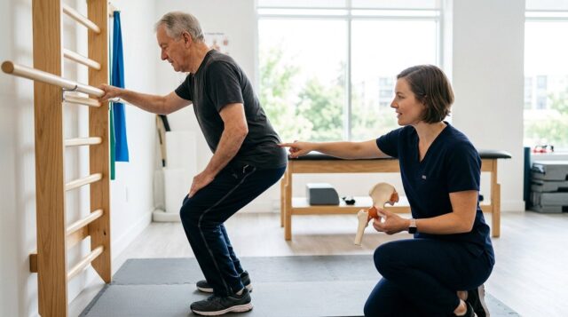
64% of patients with MRI-verified hip abductor tendon pathology reached a clinically relevant improvement in HAGOS pain after a 3-month education + exercise programme
Patients with tendon ruptures improved more than those with tendinopathy alone, though the study wasn't powered to confirm this difference
Strength gains were minimal despite clear symptomatic improvement, suggesting education and load management, not muscle capacity, may drive the benefit
Introduction
Lateral hip pain is a common and potentially disabling musculoskeletal complaint, particularly among women over 40. It is subgrouped under greater trochanteric pain syndrome (GTPS), the umbrella for lateral hip pain. Symptoms frequently originate from pathology involving the gluteus medius and/or gluteus minimus tendons at their insertion on the greater trochanter. These tendon abnormalities can range from tendinopathy to partial- or full-thickness ruptures.
Exercise therapy combined with patient education has increasingly been recommended as a first-line treatment for gluteal tendinopathy. Previous clinical trials, including the well-known LEAP trial by Mellor et al. (2018), have demonstrated encouraging outcomes with this approach.
Most of the exercise evidence we lean on comes from primary care populations with relatively mild presentations. What happens when these patients have already failed primary care and land in your secondary care caseload, with longer pain duration, more disability, and MRI-confirmed tendon damage, sometimes even ruptures? That is still unclear, as the pile of evidence is mostly derived from primary care studies. Therefore, the authors questioned whether the same education-plus-exercise approach can help those patients failing primary care and presenting in secondary care.
Methods
This was a prospective cohort study in which women between the ages of 18-75 years with lateral hip pain lasting more than 6 months were included. These participants were recruited from the orthopedic outpatient clinic. Patients were eligible when the examination revealed palpable tenderness at the insertion of the hip abductors at the greater trochanter, reproduction of known pain and/or poor pelvic stability (Trendelenburg sign) when standing on one leg, and reduced side-lying hip abduction force. Then, patients underwent MRI to verify hip abductor tendon pathology, defined as tendinopathy or rupture of gluteus medius/minimus muscles.
Patients were excluded if they had received a corticosteroid injection into the affected lateral hip within the preceding six weeks, undergone previous bone-related surgery on the affected hip, had bilateral hip abductor tendon pathology, had radiographically confirmed hip osteoarthritis (Kellgren–Lawrence grade 2 or higher), or were pregnant.
All patients received a 3-month physiotherapist-led programme in which 7 supervised education sessions (discussing load management, activity modification, and avoiding hip adduction positions) were conducted, and they received a daily home-based exercise programme consisting of 4 progressive exercises (isometric abduction, weight-bearing abduction, bridging, squats).
Outcomes
The primary outcome was the change in the Copenhagen Hip and Groin Outcome Score (HAGOS) pain subscale (0-100) at 3 months. The authors calculated the minimal important change threshold for HAGOS pain using half the baseline standard deviation.
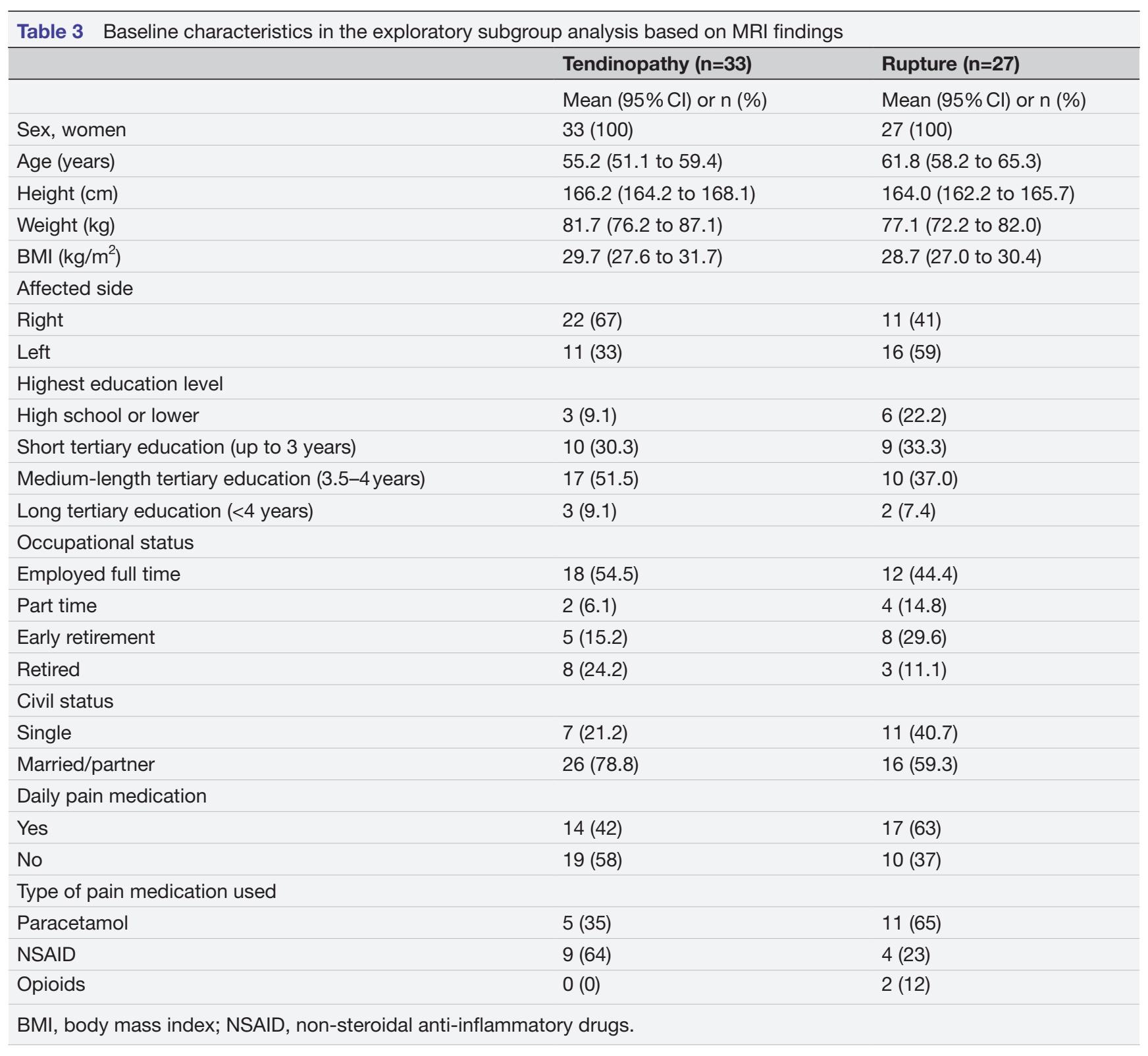
Secondary outcomes included the other HAGOS subscales, VISA-G, Oxford Hip Score (OHS), EQ-5D-5L, NRS pain, Global Rating of Change (GROC), isometric hip strength, and 30-second Chair Stand Test. An exploratory subgroup analysis compared patients with tendinopathy (n=33) vs rupture (n=27).
Results
Sixty women with MRI-confirmed hip abductor tendon pathology were included in the study, and 55 of them completed the intervention (92%). The authors reported 85% exercise adherence and zero adverse events.
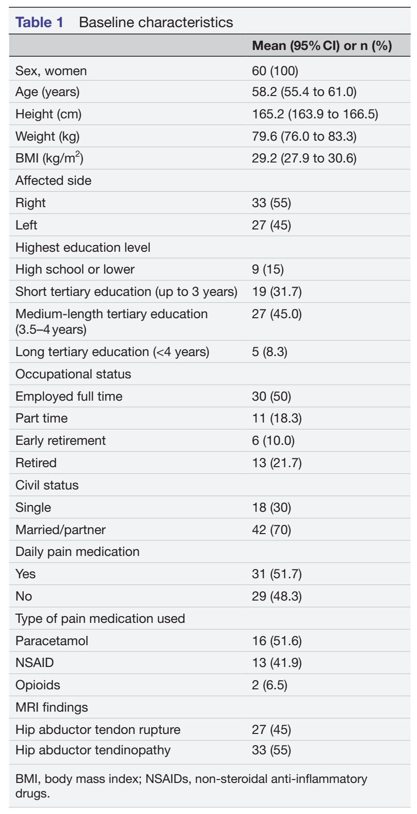
The primary outcome, HAGOS pain, improved from 46.4 to 60.1 points (mean change 14.5, 95% CI 9.0 to 20.0), and 64% of patients exceeded the minimal important change of 9.3 points.
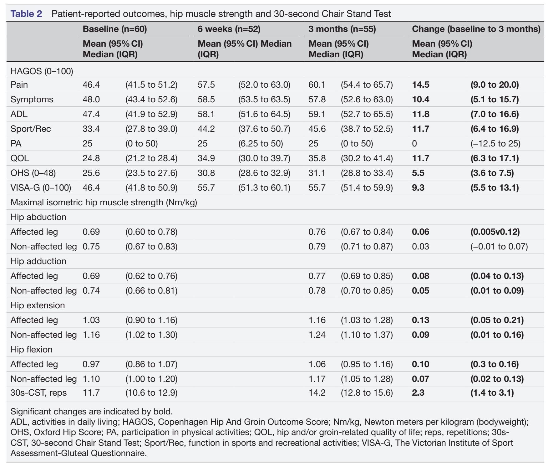
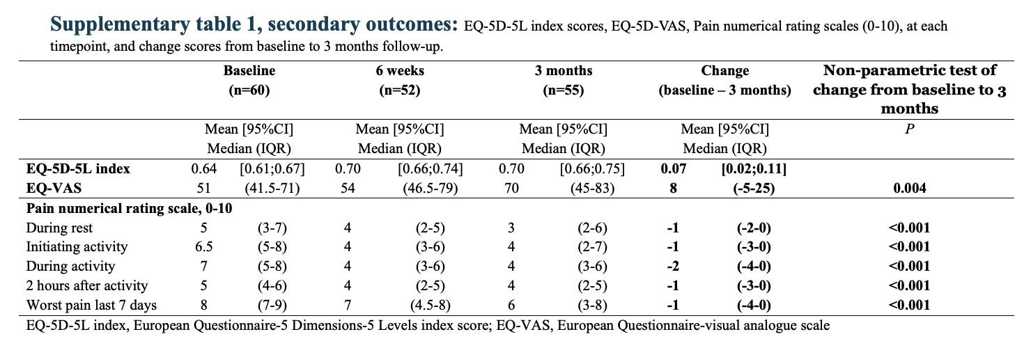
Nearly all secondary patient-reported outcomes improved significantly (VISA-G +9.3, OHS +5.5, EQ-5D-5L and NRS pain measures), except the HAGOS physical activities subscale, which didn’t budge.
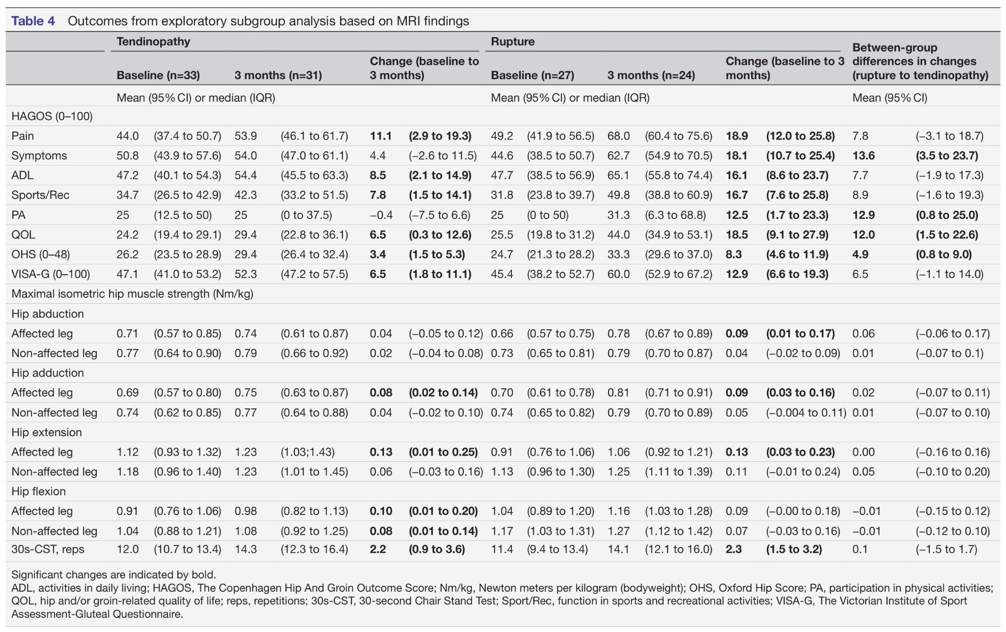
Functionally, the 30-second Chair Stand Test improved from 11.7 to 14.2 repetitions, but isometric hip strength gains were small across nearly all directions. On the Global Rating of Change, only 37% reported successful improvement at 3 months.
In the exploratory subgroup analysis, the rupture group (n=27) improved more on HAGOS pain (+18.9 points) than the tendinopathy group (n=33, +11.1 points), and a successful GRoC was reported by 50% of the rupture group vs. 26% of the tendinopathy group. However, the between-group difference wasn’t statistically significant, and the study wasn’t powered for this comparison.
Questions and thoughts
Stronger means better, right?
The most clinically interesting finding here might be the mismatch between strength and symptom improvement. We tend to assume that if someone’s hip hurts less and moves better, their muscles must have gotten stronger. This study, like Mellor et al.’s earlier RCT, shows that’s not necessarily true: strength gains were minimal to negligible, yet pain and function clearly improved. This fits a growing body of evidence that education, addressing fear of loading, optimising daily movement strategies, and improving adherence may be doing more heavy lifting than the exercises themselves in terms of mechanism of improvement. That doesn’t mean exercise is irrelevant, but it reframes what we might be achieving with it: not necessarily “strengthening” the abductors, but building tolerance to load and confidence in using the hip again.
Why did so few patients report an overall successful improvement?
The authors further report that 64% of the participants reached the minimal important change of 9.3 points, which was defined as half the standard deviation of the baseline scores. Strikingly, only 37% of the participants reported a successful overall improvement on the GRoC. Instead of framing it as two-thirds of the cohort improving successfully, saying that only slightly more than a third of the participants improved sounds more rational. The study did not investigate individual goals or reasons for patients’ GRoC responses. This highlights a potential issue in rehabilitation assessment: improvement in a symptom-specific questionnaire may not adequately represent a patient’s overall perception of recovery. So, monitoring meaningful activities and participation alongside pain questionnaires may therefore be valuable in clinic settings, although this particular study did not test such a strategy.
Could age influence the underlying pathology and response to rehabilitation?
The study included patients aged 18–75 years, representing a potentially broad range of underlying musculoskeletal conditions. In younger patients, conditions such as femoroacetabular impingement (FAI), hip dysplasia, or musculotendinous injuries may contribute to hip pain, whereas older patients may be more likely to present with degenerative tendon changes, fatty infiltration, muscle atrophy, or coexisting joint pathology. Although radiographically confirmed hip osteoarthritis was excluded, other potentially relevant hip conditions were not explicitly addressed in the exclusion criteria.
Importantly, all participants had MRI-confirmed hip abductor tendon pathology, but the presence of structural abnormalities does not necessarily establish that these findings were the sole source of symptoms. Different age groups may therefore have different contributing mechanisms and potentially different responses to the same rehabilitation programme.
The authors did not investigate whether age influenced treatment outcomes. This raises the question of whether a standardised education and exercise approach is equally appropriate across the entire age spectrum, or whether younger and older patients require different rehabilitation considerations.
Can patients with MRI-confirmed gluteal tendon rupture be treated conservatively?
The subgroup finding (bigger gains in the rupture group) is intriguing but needs to be read with caution. It’s exploratory, underpowered, and purely descriptive. Still, it pushes back against the instinct many clinicians (and surgeons) have that a confirmed tendon rupture automatically means “this needs surgery.” If anything, these ruptured tendons responded just as well, if not better, to conservative management than the tendinopathy group did, BUT this is exploratory and should be further investigated first.
Worth noting too: these are patients who had already failed to improve enough in primary care, yet a relatively modest programme (7 sessions over 3 months) still let them improve. That’s a strong argument for exhausting conservative, physiotherapist-led care even in patients who’ve been bounced up to secondary/orthopaedic settings, before jumping to surgical reconstruction discussions.
Talk nerdy to me
This was a single-arm cohort study with no control group, so we cannot attribute the improvements to the intervention with certainty. The authors are upfront about this: natural history data for gluteal tendinopathy shows that even without structured treatment, 29-52% of patients can report successful improvement on GRoC within a year. But they balance this by citing an 11-year natural history study where 45% remained symptomatic, suggesting these secondary-care patients, with longer symptom duration and higher baseline disability, were unlikely to resolve spontaneously within 3 months.
Furthermore, the combined intervention design also means education and exercise can’t be disentangled. We don’t know how much of the effect comes from the exercises versus the education and load-management advice, something the authors flag as a priority for future research. Similarly, self-efficacy, a known mediator of outcome in this population, was not measured, but the 2022 Mellor paper already found that self-efficacy was an important mediator, whereas strength and range of motion improvements were not.
Moreover, four standard exercises were provided, although with different progression levels. This is doable in a study environment, yet, in clinical practice we won’t give the same exercises to different people with different goals. Individualizing and adapting the exercises according to the needs of the person in front of you remains key.
The subgroup classification into tendinopathy vs. rupture was done by a single evaluator using a locally developed MRI reading protocol, applied after the intervention was already completed (but blinded to outcome), which limits how confidently we can read into that comparison. Combined with the lack of statistical power for between-group testing, treat those numbers as hypothesis-generating only, not as evidence that ruptures respond better than tendinopathies.
Finally, this is a single-centre Danish cohort, 100% women, which limits generalisability, though that’s consistent with the epidemiology of this condition.
Take-home messages
Following a three-month physiotherapist-led education and exercise programme, approximately two-thirds of secondary-care patients with MRI-confirmed hip abductor tendon pathology, including tendon ruptures, achieved a clinically relevant improvement in pain. However, only 37% reported a successful overall improvement on the Global Rating of Change (GRoC), suggesting that reduced pain did not necessarily translate into a satisfactory recovery. The relatively small strength gains suggest that improvements in symptoms may not depend primarily on increased muscle strength, although the contribution of education, load management and exercise remains uncertain. These findings are encouraging for conservative management, even in patients with tendon ruptures, but the absence of a control group prevents attributing the improvements directly to the intervention or establishing its effectiveness as an alternative to surgery.
Reference

