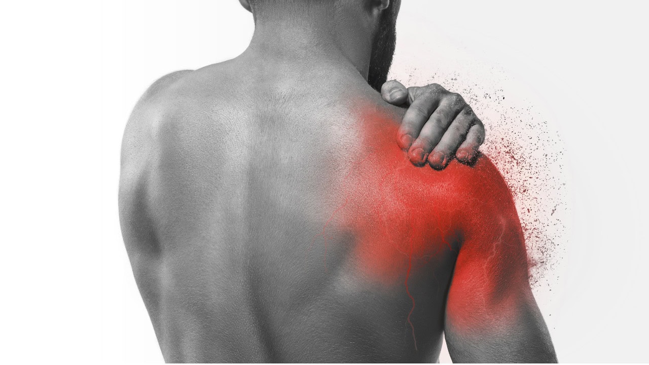

Glenohumeral instability describes symptomatic excessive translation of the humeral head relative to the glenoid. It may present as apprehension, pain, a sensation of slipping or giving way, subluxation, or complete dislocation. Importantly, laxity alone is not instability: a patient may have considerable passive translation without symptoms when dynamic muscular control is sufficient.
Epidemiology and prognosis
Approximately 75% of shoulder instability presentations are traumatic, around 75% occur predominantly in an anterior direction, 20% posteriorly and approximately 5% are multidirectional, commonly in association with hypermobility. Anterior instability refers to excessive anterior translation of the humeral head relative to the glenoid; posterior instability describes excessive posterior translation.
Traumatic anterior dislocation typically occurs with forced abduction and external rotation. Associated pathology is common: Bankart lesions have been reported in 84–97% of anterior dislocations and Hill-Sachs lesions in 41–83%. Age modifies the associated injury pattern. In younger patients, isolated traumatic cuff tears are relatively uncommon, whereas recurrence is an important concern, particularly in young contact athletes. With increasing age, traumatic rotator cuff tears, greater tuberosity fractures and neurological injuries become increasingly relevant following dislocation.
For atraumatic and multidirectional instability, physiotherapy remains first-line management. The Watson rehabilitation programme is typically delivered over approximately 3–6 months, although more complex persistent instability may require considerably longer rehabilitation.
Do not equate multidirectional laxity with multidirectional symptomatic instability.
1. Screening and History
A thorough history should extend beyond the most recent episode. Ask about previous apprehension, subluxation, complete dislocation, voluntary displacement, trauma and generalized joint laxity.
Age and mechanism
The mechanism and patient profile help estimate what may be driving the presentation. A young collision athlete with a high-force dislocation raises concern for recurrent traumatic instability and structural damage. After traumatic dislocation in an older patient, the probability of associated cuff tear, greater tuberosity fracture or neurological injury becomes more important. These are not age-exclusive diagnoses, but their relative likelihood changes across age groups.
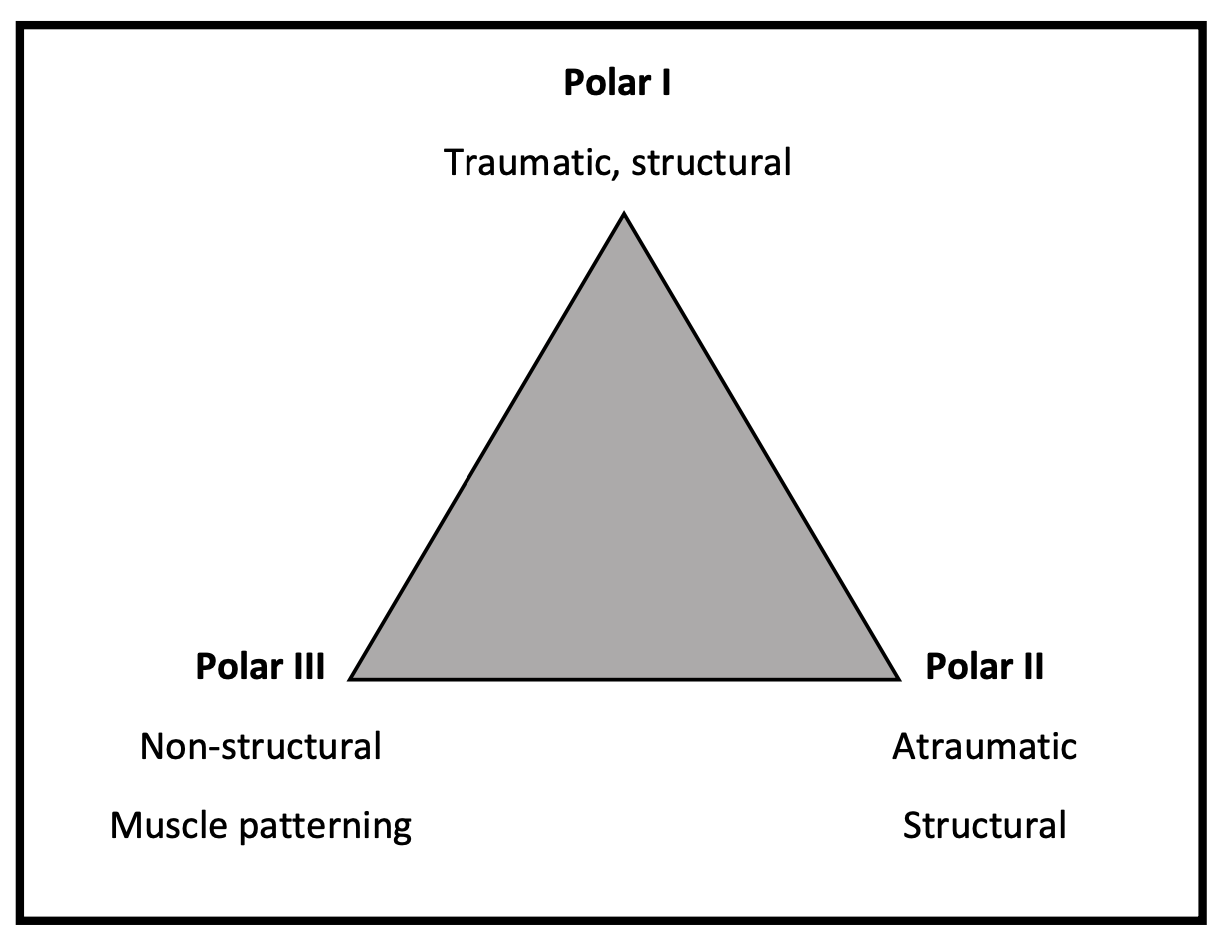
The history can also help position the patient along the Stanmore instability continuum:
- Polar I: traumatic with structural pathology;
- Polar II: atraumatic with a structural component;
- Polar III: predominantly non-structural muscle-patterning.
Rather than viewing these as rigid diagnostic boxes, use them to understand what proportion of the patient’s presentation appears structural versus motor-control related. A patient close to Polar I may require surgical assessment when substantial structural pathology is present. As the presentation moves toward Polar III, rehabilitation aimed at neuromuscular control becomes increasingly central. Polar II often sits between these extremes, with structural and motor-control factors both contributing.
Clarify the type of instability
Determine:
- the symptomatic direction: anterior, posterior, inferior or more than one direction;
- subluxation versus complete dislocation;
- first versus recurrent instability episode;
- whether progressively less force is required to provoke symptoms;
- whether instability occurs during daily activities or sleep.
Do not equate multidirectional laxity with multidirectional symptomatic instability. A patient may be lax in several directions but experience apprehension in only one or two.
Also ask about deliberate “party trick” displacement, particularly where a muscle-patterning presentation is suspected.
Generalized hypermobility
Assess for excessive laxity elsewhere and, where indicated, use the Beighton score. Generalized hypermobility may indicate that the shoulder presentation is part of a broader hypermobility spectrum rather than an isolated glenohumeral problem.
Fear and confidence
Ask which movements the patient avoids, whether they trust the shoulder and which sporting situations provoke apprehension. Fear, anxiety, avoidance and low self-efficacy may influence persistent atraumatic instability and later return to sport.
After traumatic anterior dislocation: operative or non-operative rehabilitation?
The 2026 British Elbow and Shoulder Society (BESS) practice guideline provides a unified rehabilitation framework for adults with traumatic anterior shoulder instability managed either non-operatively following closed reduction or post-operatively following arthroscopic Bankart repair.
Both pathways are organized around five sequential phases:
- Acute protection and education
- Movement and early strengthening
- Progressive strengthening
- Return to sport
- High-level function
The first three phases broadly correspond to approximately 0–3, 3–6 and 6–12 weeks, with return-to-sport work beginning after approximately 12 weeks. However, BESS emphasizes that later progression should increasingly depend on functional status, confidence, absence of apprehension and psychological readiness, rather than time alone.
Following arthroscopic Bankart repair, current BESS consensus supports approximately 2–3 weeks of sling use and delaying through-range strengthening until around 6 weeks. Non-operative rehabilitation follows a similar overall structure but allows earlier sling weaning. These recommendations are pragmatic consensus guidance rather than high-certainty evidence.
Where the history or examination raises concern for fracture, substantial bone loss, traumatic cuff injury or neurological involvement, further medical assessment remains appropriate. The BESS guideline itself is a rehabilitation framework rather than a complete imaging or referral algorithm, so these decisions still need to follow local trauma and orthopaedic pathways.
2. Physical assessment: identify the dominant contributor
The purpose of examination is not simply to confirm instability but to determine what is driving the patient’s symptoms.
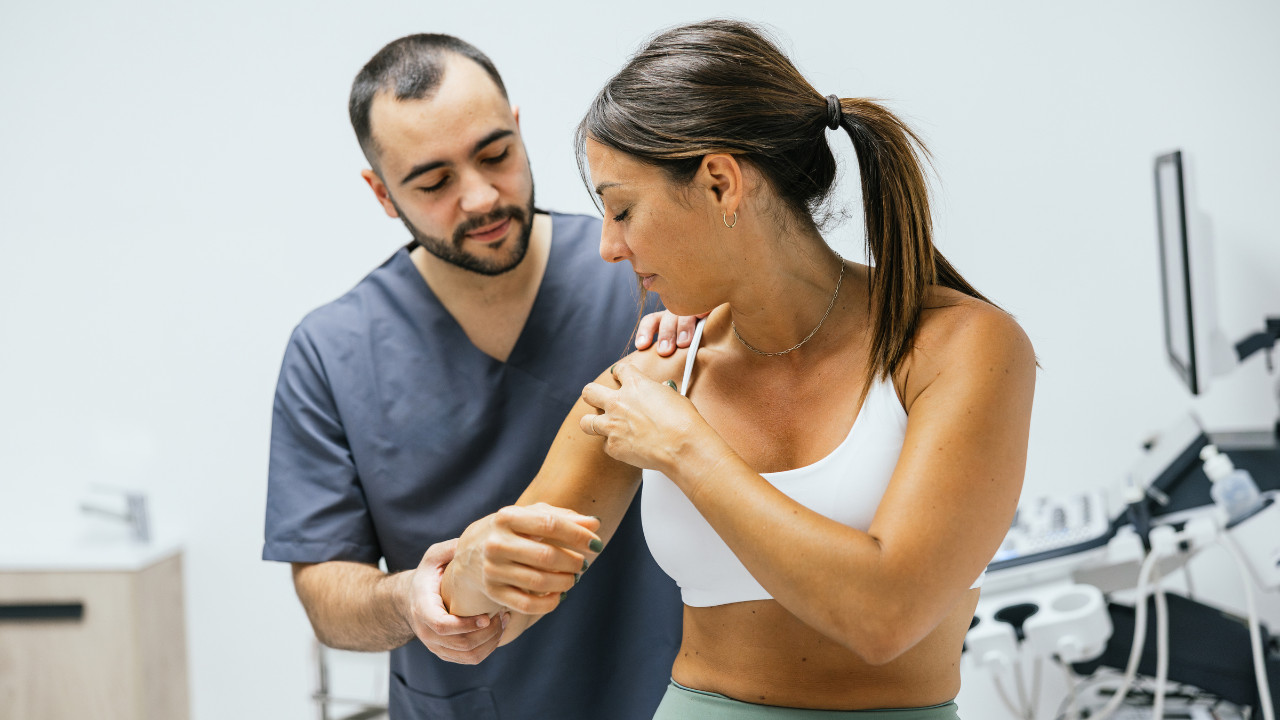
Look, feel and move
Observe posture and resting scapular position, including medial-border or inferior-angle prominence, protraction and downward rotation. A relatively downwardly rotated scapula has been described particularly in inferior, posteroinferior and multidirectional instability.
Palpate the clavicle, AC joint, acromion, coracoid, scapular spine and anterior/posterior glenohumeral joint lines. This helps identify local tenderness and possible associated pathology. Increased resting tone in upper trapezius, pectoralis major or latissimus dorsi may also suggest altered recruitment strategies.
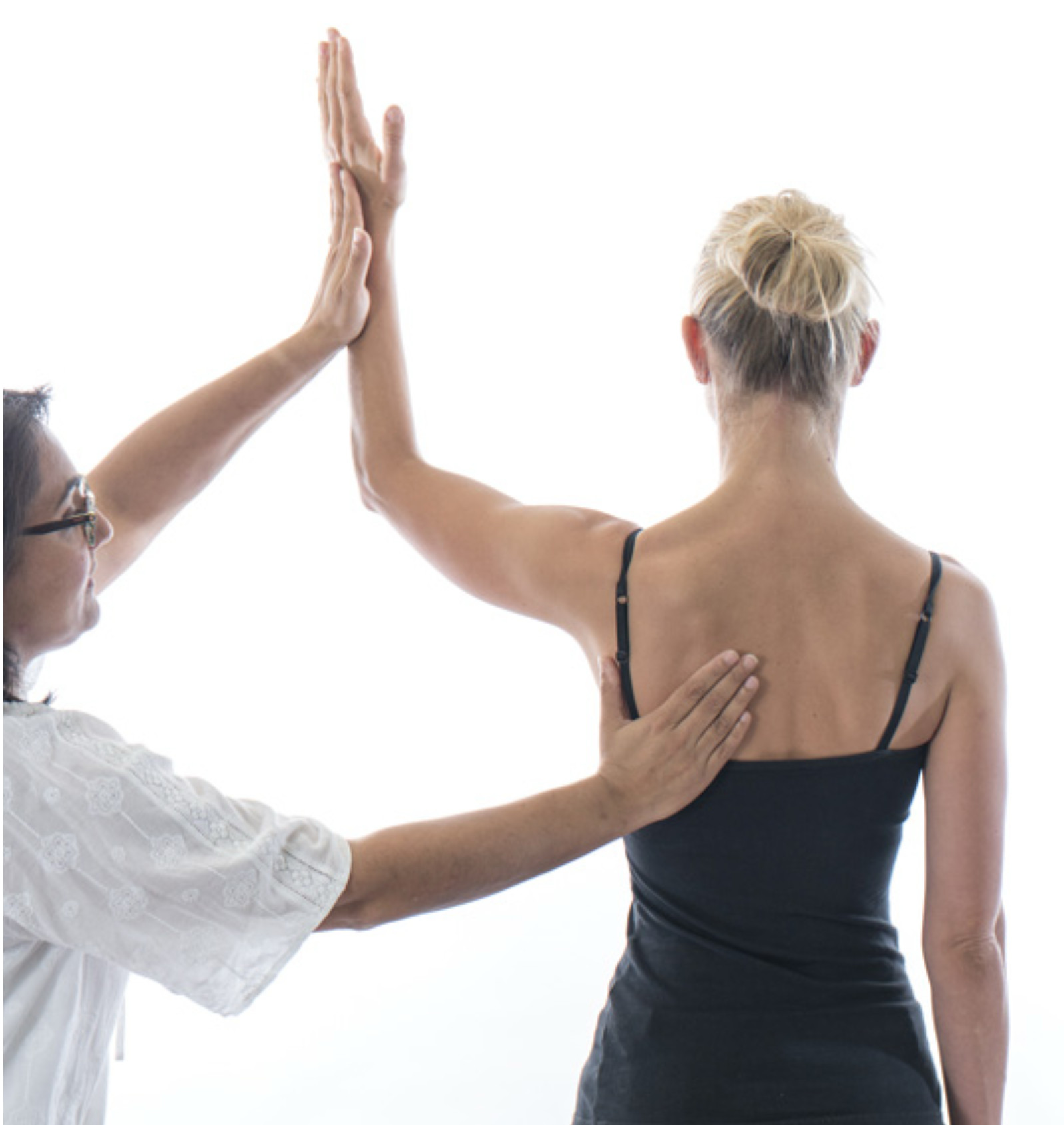
Assess active scapular elevation/depression and protraction/retraction. If movement appears poorly controlled, repeat it while supporting the arm. If scapular control improves when the weight of the arm is supported, the abnormal scapular movement may be secondary to insufficient shoulder control under load rather than a primary scapular impairment.
Then observe at least three repetitions of bilateral forward flexion and abduction, looking for range, fatigue, pain, apprehension and scapulothoracic control.
Assess internal and external rotation with the elbow flexed to 90°:
- with the humerus by the side;
- at 90° shoulder abduction.
Test the cuff through inner and outer range
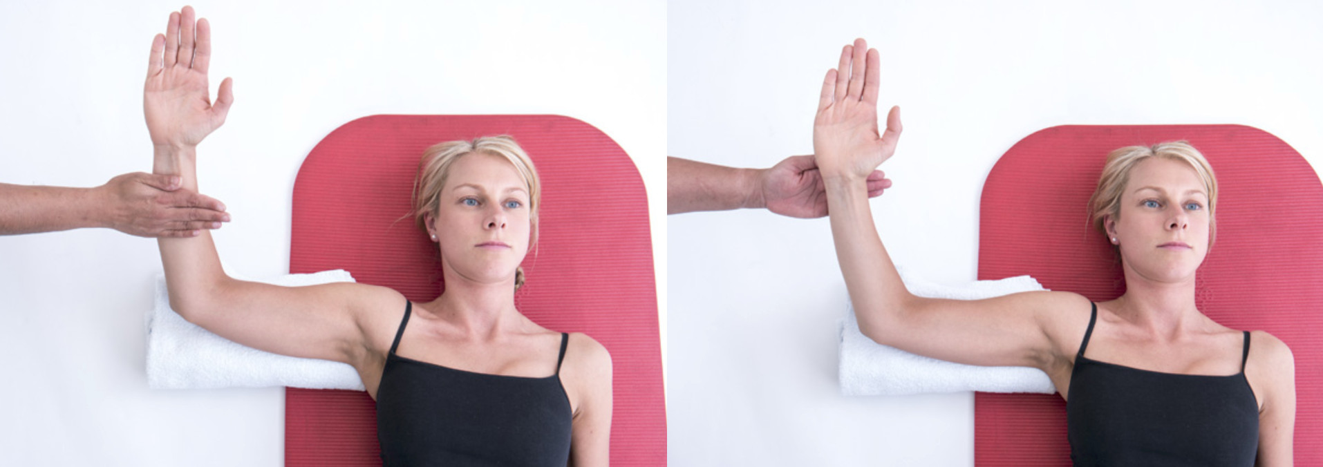
Position the patient supine with:
- shoulder abducted 90°;
- elbow flexed 90°;
- a small rolled towel beneath the humerus to support the arm weight and prevent glenohumeral extension.
At maximum external rotation:
- resisted ER tests posterior cuff in inner range;
- resisted IR tests anterior cuff in outer range.
At maximum internal rotation:
- resisted ER tests posterior cuff in outer range;
- resisted IR tests anterior cuff in inner range.
This can reveal range-specific deficits that a single mid-range strength measure may miss.
Screen the kinetic chain
Lower-limb and trunk deficits may alter force transfer to the upper limb. One option is the corkscrew test: ask the patient to perform a single-leg squat and observe excessive rotational collapse through the hip and knee. If this happens, exercises to improve lower limb and or core stability should be included in the early phases of rehabilitation.
Symptom-modification tests
Jaggi and Alexander recommend repeating symptomatic forward flexion or abduction while modifying:
- upright posture/scapular position;
- kinetic-chain contribution by rising onto the toes;
- scapular position manually;
- cuff/deltoid activity using external-rotation resistance.
If one modification improves symptoms, range or control, investigate that component further and consider incorporating it into rehabilitation.
Specific instability tests
Anterior instability
To assess anterior instability, combining the Apprehension and Relocation Tests has been reported to provide 67% sensitivity and 98% specificity, making a positive combined finding useful for ruling in anterior structural instability (Hegedus et al., 2012).
The apprehension test should be followed by the relocation test.
Posterior instability
For posteroinferior instability, the Jerk Test can be useful, with reported sensitivity of 90% and specificity of 85% (Kim et al., 2004). The nature of the positive response may also have prognostic value: a painful Jerk Test was associated with poorer outcomes after conservative care, whereas 93% of patients with a painless clunk improved with rehabilitation over a mean period of about 4 months.
Inferior instability
Inferior glenohumeral laxity can be assessed with the Sulcus Sign and Gagey hyperabduction test, although neither should be used in isolation. A Sulcus Sign of >2 cm has been reported to have 28% sensitivity and 97% specificity for multidirectional instability, while Eshøj et al. found only moderate inter-rater reliability (κ = 0.43) (Tzannes & Murrell, 2002; Eshøj et al., 2018). More recently, the Gagey test showed 46% sensitivity and 38% specificity for detecting inferior glenohumeral ligament lesions, supporting its use as an adjunct rather than a stand-alone diagnostic test (van Spanning et al., 2023).
3. Treatment: progress rehabilitation by increasing task demand
Shoulder instability rehabilitation should not be treated as a fixed sequence of exercises. Across the available rehabilitation frameworks, however, there is a common clinical logic: begin at a level where the patient can control the shoulder, then progressively increase the demands of range, load, speed, reactivity and sport specificity. These domains can overlap, and the starting point will depend on the instability phenotype, symptom irritability and deficits identified during assessment.
Establish a controllable starting point
If the patient cannot control the shoulder comfortably against the weight of the arm, reduce the demand first.
Jaggi and Alexander describe using supported positions such as supine, prone or sitting with the arm supported on a table, the patient’s thigh or towels. Closed-chain positions against a wall, table or floor can also facilitate rotator cuff and deltoid co-contraction while reducing apprehension.
For poor rotational control, internal and external rotation can initially be practised with the arm supported. Progress by increasing the available rotational arc, reducing support, increasing shoulder abduction and eventually adding external resistance.
For anterior instability with an inner-range external-rotation deficit, Jaggi and Alexander give a specific example:
prone, arm fully supported, inner-range external rotation:
8–10 repetitions × 2–3 sets, twice daily.
Restore control through increasing range
Once the patient can control the shoulder in lower-demand positions, progressively challenge control through larger ranges.
The Watson programme illustrates this particularly well in multidirectional instability. Scapular upward-rotation control is initially trained in standing at approximately 20–30° abduction, before progressing glenohumeral control through approximately 0–45° abduction, then toward 90° and eventually higher functional ranges.
Examples include:
- scapular upward-rotation drill at 20–30° abduction: 1–3 sets × 20 repetitions, 1–2 times/day;
- ER/IR or extension isometrics at low abduction angles: 5–10 × 5-second holds at approximately 20–30% MVC, 2–3 times/day;
- band-resisted ER, IR or extension in the 0–45° range: 1–3 sets × 20 repetitions, twice daily;
- side-lying external rotation: 1–3 sets × 20 repetitions, twice daily, progressing from arm weight to approximately 0.5–1 kg initially.
Forward-flexion drills then progress from approximately 20–30° toward 45° and 90°, while ER and IR can later be trained at 90° abduction once adequate scapular and humeral-head control has been established.
Progression should depend on maintaining satisfactory scapular and humeral-head control and keeping symptoms acceptable, rather than simply reaching a predetermined time point.
Develop endurance
Once the patient can reproduce the desired movement strategy, the next challenge may be sustaining it under repeated loading and fatigue.
In the Watson programme, an endurance dosage is suggested for the scapular, rotator cuff and deltoid exercises already established earlier in rehabilitation:
1–3 sets × 10–15 repetitions, 1–2 times/day.
This dosage is not tied to one single exercise; it is a loading progression for exercises such as resisted ER/IR, scapular-control drills, forward flexion or rowing once the primary aim shifts from motor recruitment toward endurance.
Develop strength
As movement becomes more robust, increase external resistance and reduce repetition volume.
For established rotator cuff, scapular and deltoid strengthening exercises, Watson et al. suggest progressing toward:
3–4 sets × 8–12 repetitions, every second day.
Resistance can be progressed with heavier bands or external weights according to the patient’s functional requirements. Strength should be developed in the ranges relevant to the instability presentation rather than only with the arm by the side.
Add speed, deceleration and reactive stability
Slow strength does not fully prepare an athlete for rapid perturbations, catching, contact or force absorption.
The Derby Shoulder Instability Programme introduces faster muscle activation, plyometrics and deceleration using exercises such as:
- 1-kg drop-and-catch at 90° scaption: target 100 repetitions;
- progress to contralateral single-leg stance or eyes closed: target 100 repetitions;
- standing falling press-up: 50 repetitions;
- waist-height falling press-up: 50 repetitions;
- plyometric push-up with hand clap: 20 repetitions;
- doorway fall: 20 repetitions.
Its proprioceptive and closed-chain section progresses exercises such as single-hand ball rolling, kneeling weight shifts and push-up-position variations toward 60-second targets, performed twice daily.
These are progression targets from the Derby programme, not universal prescriptions for every patient with instability.
Online Course
Learn advanced, evidence-based strategies to assess sporting shoulder injuries, build individualized rehabilitation programmes, and guide athletes safely from diagnosis through return to play with Edel Fanning, PhD.

Progress to sport-specific part practice
Once adequate control and capacity are present, rehabilitation should increasingly resemble the specific demands of the patient’s sport.
Watson et al. describe part practice: breaking a complex movement into components before reintegrating the complete task. For example, a swimmer may separately practise the catch, pull and recovery phases before returning to the complete stroke. With overhead athletes, the Physiotutors Shoulder Rehabilitation Course with Edel Fanning emphasizes end-range rotational control, posterior cuff loading, plyometric ball work, throwing progression, thoracic rotation and kinetic-chain integration. For contact athletes, relevant preparation may include:
bear crawls → grappling → controlled falls → progressive contact → tackling preparation.
Reintegrate whole practice
Once the difficult components can be performed with adequate control, progress toward the complete sporting or occupational task.
Watson et al. recommend increasing from part practice to whole practice, with training volume gradually increased toward the demands of the patient’s sport or occupation.
At this stage, rehabilitation should progressively reproduce the required range, load, speed and unpredictability of the activity.
Return to sport
Return-to-sport decisions should consider more than time since injury or surgery.
Assess, where relevant:
- range-specific rotator cuff strength;
- explosive and functional upper-limb performance;
- open- and closed-chain capacity;
- tolerance of sport-specific exposure;
- absence of meaningful apprehension;
- psychological readiness.
The Shoulder Instability Return to Sport after Injury questionnaire (SIRSI) is a patient-reported measure of psychological readiness. A SIRSI score ≥55 has been proposed as a useful benchmark after shoulder stabilization surgery, while approximately 51.5 has shown some ability to discriminate recurrence risk. These cut-offs come from postoperative athletic cohorts and should not be used as stand-alone clearance criteria or automatically generalized to non-operative or atraumatic instability.
For traumatic anterior instability, the BESS guideline similarly emphasizes that later rehabilitation and return-to-sport decisions should increasingly be guided by function, confidence, absence of apprehension and psychological readiness, rather than time alone.
Clinical takeaway
Shoulder instability rehabilitation should be individualized to the instability phenotype and the deficits identified during assessment, rather than following one fixed exercise protocol. Across the different rehabilitation frameworks, the common principle is to begin with a level of demand the patient can control, then progressively challenge range, endurance, strength, speed, reactivity and sport-specific function as appropriate.
The key is not whether a patient completes a particular exercise, but whether they can maintain satisfactory glenohumeral and scapular control as the task becomes more demanding. Return to sport should therefore be based on a combination of physical capacity, sport-specific exposure, absence of meaningful apprehension and psychological readiness, rather than on time alone.
References
Bateman, M. (2013). Derby Shoulder Instability Rehabilitation Programme (Version 1.0) [Rehabilitation protocol].
Eshoj, H., Ingwersen, K. G., Larsen, C. M., Kjaer, B. H., & Juul-Kristensen, B. (2018). Intertester reliability of clinical shoulder instability and laxity tests in subjects with and without self-reported shoulder problems. BMJ Open, 8(3), e018472. https://doi.org/10.1136/bmjopen-2017-018472
Hegedus, E. J., Goode, A. P., Cook, C. E., Michener, L., Myer, C. A., Myer, D. M., & Wright, A. A. (2012). Which physical examination tests provide clinicians with the most value when examining the shoulder? Update of a systematic review with meta-analysis of individual tests. British Journal of Sports Medicine, 46(14), 964–978. https://doi.org/10.1136/bjsports-2012-091066
Jaggi, A., & Alexander, S. (2017). Rehabilitation for shoulder instability—Current approaches. The Open Orthopaedics Journal, 11, 957–971. https://doi.org/10.2174/1874325001711010957
Kim, S.-H., Park, J.-C., Park, J.-S., & Oh, I. (2004). Painful jerk test: A predictor of success in nonoperative treatment of posteroinferior instability of the shoulder. The American Journal of Sports Medicine, 32(8), 1849–1855. https://doi.org/10.1177/0363546504265263
Pasqualini, I., Hurley, E. T., Khan, S. T., Soares, R. W., Grobaty, L., Johnson, C., Lau, B. C., Tjong, V. K., & Rossi, L. A. (2025). Psychological readiness for return to sport after shoulder stabilization surgery: A review of current evidence and the role of the Shoulder Instability Return to Sport After Injury (SIRSI) scale. Open Access Journal of Sports Medicine, 16, 55–65. https://doi.org/10.2147/OAJSM.S505455
Pasqualini, I., Rossi, L. A., Hurley, E. T., Turan, O., Tanoira, I., & Ranalletta, M. (2024). Shoulder Instability-Return to Sports After Injury scale shows that lack of psychological readiness predicts outcomes and recurrence following surgical stabilization. Arthroscopy: The Journal of Arthroscopic & Related Surgery, 40(12), 2815–2824. https://doi.org/10.1016/j.arthro.2024.04.030
Pavlenco, C., Khilfeh, B., Wang, X., & Saper, M. (2026). Six-month SIRSI scores and return to sport after shoulder stabilization in adolescent athletes. Orthopaedic Journal of Sports Medicine, 14(3), 23259671261421228. https://doi.org/10.1177/23259671261421228
Tzannes, A., & Murrell, G. A. C. (2002). Clinical examination of the unstable shoulder. Sports Medicine, 32(7), 447–457. https://doi.org/10.2165/00007256-200232070-00004
van Spanning, S. H., Lafosse, T., Verweij, L. P. E., van Rijn, S. K., Lafosse, L., & Buijze, G. A. (2023). Predictive value of Gagey’s hyperabduction test in identifying inferior glenohumeral ligament lesions. Orthopaedics & Traumatology: Surgery & Research, 109(4), 103500. https://doi.org/10.1016/j.otsr.2022.103500
Watson, L., Warby, S., Balster, S., Lenssen, R., & Pizzari, T. (2016). The treatment of multidirectional instability of the shoulder with a rehabilitation program: Part 1. Shoulder & Elbow, 8(4), 271–278. https://doi.org/10.1177/1758573216652086
Watson, L., Warby, S., Balster, S., Lenssen, R., & Pizzari, T. (2017). The treatment of multidirectional instability of the shoulder with a rehabilitation programme: Part 2. Shoulder & Elbow, 9(1), 46–53. https://doi.org/10.1177/1758573216652087
Wong, C., Jaggi, A., Kearney, R., & Gwilym, S. (2026). British Elbow and Shoulder Society practice guidelines: Rehabilitation following traumatic anterior shoulder dislocation (post-operative and non-operative care). Shoulder & Elbow, 18(3), 604–608. https://doi.org/10.1177/17585732261439731