Dizziness & Vertigo

BPPV / Benign Paroxysmal Positional Vertigo | Diagnosis & Treatment

Check our shop
Bppv

BPPV / Benign Paroxysmal Positional Vertigo | Diagnosis & Treatment

Benign paroxysmal positional vertigo, abbreviated as BPPV is the most common inner ear problem and cause of vertigo, or a false sense of spinning. Common causes are head trauma or ear infections, although most cases appear to be idiopathic.BPPV can be caused by debris in the semicircular canal of the ear, which continues to move after the head has stopped moving. This causes ongoing movement that conflicts with other sensory information.

Right inner ear

The semicircular canals are filled with a fluid called endolymph. The main sense organ in each canal is called the crista, which is stimulated by the movement of the cupula. Head rotation causes relative movement of the endolymph in the semicircular canal, which bends the cupula and the embedded hairs of the hair cells and causes stimulation of the relevant vestibular nerve. The cause of BPPV is believed to be canalithiasis, affecting the posterior semicircular canal in 85 to 95% of all cases. In canalithiasis, free-floating debris in the semicircular canal is hypothesized to act like a plunger, causing continuing movement of the endolymph even after head movement has ceased. This causes movement of the cupula and bending of the hairs of the hair cells and provokes vertigo.

Endolymph

Around 20% of BPPV cases are said to resolve within 4 weeks and up to 50% up to 3 months without treatment, but recurrence is reported between 10-18% after 1 year.

Like what you're learning?

Follow a course

  • Learn from wherever, whenever, and at your own pace
  • Interactive online courses from an award-winning team
  • CEU/CPD accreditation in the Netherlands, Belgium, US & UK

Clinical Picture

It can be helpful to recall that vertigo requires an imbalance between the two sides (Molnar et al. 2014). Initially, clinicians should categorize the patient’s dizziness into 1 of the following 3 types:
1) Vertigo
2) Light-headedness
3) Disequilibrium

This approach is based on early investigations of chronic dizziness (Drachman et al. 1972). The following table shows the three main types of dizziness including their specific underlying diseases:

Vertigo is a sensation of movement such as twirling or tilting perceived in the head, a symptom implying either peripheral or central vestibular disorder. All vertigo is abrupt in onset, episodic, and aggravated by head movements. Different types can be distinguished by duration, setting, and associated symptoms (Molnar et al. 2014).
Light-headedness is a feeling of faintness or graying of vision, implying hypotension and poor perfusion of the brain. Disequilibrium is a sensation of unsteadiness not in the head, implying proprioceptive or cerebellar disease (Molnar et al. 2014). However, a substantial overlap of dizziness types exists (Kerber et al. 2017).
The following questions can be helpful to further distinguish between the 3 main types of dizziness:

Vertigo patient history


Examination

Posterior Canal BPPV

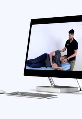
The Dix-Hallpike maneuver is considered the gold standard for the diagnosis of posterior canal BPPV. The lack of alternative external gold standards limits the availability of sensitivity and specificity data. As the Dix-Hallpike maneuver is the best test we have and is regarded as the gold standard, we are giving this test a high clinical value in practice.
Before you conduct the test, the patient should be counseled that his symptoms of vertigo will be reproduced and that he might feel nauseous. So make sure you have a bucket at hand, in case your patient might need it.

To perform the Dix-Hallpike test have your patient sit on the treatment bench in long-sit with a pillow on the table that will make sure that the patient’s head is extended to 20° in a second. Stand on the side to be examined and firmly hold your patient’s head in 45 degrees of rotation towards the side to be tested. In this case, the left posterior semicircular canal of the patient is aligned with the Sagittal plane. Instruct the patient to keep his eyes open and take the patient backward in a quick movement so that your patient’s head is still rotated and extended to 20° by the pillow.

Observe the patient’s eyes for the latency, duration, and direction of the nystagmus. The nystagmus typically usually has a latency of around 5-20 seconds and fatigues within 60 seconds after onset. In a positive test, the patient will experience vertigo during this test.
In the case of posterior canal BPPV, the nystagmus will be upbeating and torsional, meaning that the upper pole of the eye is beating toward the dependent ear, and the vertical component beating toward the forehead.

After the resolution of subjective vertigo and nystagmus, if present, the patient may be slowly returned to the upright position. The nystagmus might be seen again in the reverse direction after the patient returns to the upright position and should be allowed to resolve. If the initial result is negative, the Dix-Hallpike test should be repeated for the other side.

If the nystagmus presented with a lateral beat or a downbeat, lateral or anterior BPPV should be suspected. On top of that, If you suspect BPPV in your patient and this maneuver is negative in both directions, you should assess the lateral canal with the Supine Head Roll maneuver. The anterior canal is rarely affected with 1-3% of all BPPV cases and its pathophysiology is poorly understood. In these cases, you should refer to a specialist.


Lateral Canal BPPV

To perform the Supine Head Roll test have your patient lie supine on the treatment bench and bring his head into 30 degrees of flexion to align the lateral semicircular canal in the horizontal plane. Then quickly rotate 90 degrees to one side and observe your patient’s eyes for nystagmus, which usually has a latency of 5-20 seconds and fatigues within 60 seconds after onset. After the nystagmus subsides (or if no nystagmus is elicited), the head is then returned to the straight faceup supine position. After additional elicited nystagmus has subsided, the head is then quickly turned 90 degrees to the opposite side and the eyes are once again observed for nystagmus

In a positive test, the patient will experience vertigo during this test. In the case of lateral semicircular canal BPPV, the nystagmus will be predominantly horizontal. Two potential nystagmus findings might occur:

  • Geotropic type of nystagmus is marked by very intense horizontal nystagmus beating towards the earth on the affected side and usually less intense beating to the earth on the healthy side. It seems probable that in this form of nystagmus, the calcium carbonate debris is located in the long arm of the semicircular canal
  • Apogeotropic type: Less common with a horizontal nystagmus beating towards the uppermost ear on both sides. In this case, it is reasoned that the calcium carbonate debris is located adherently or close to the ampulla of the semicircular canal. In this case, the side opposite the strongest nystagmus is the affected ear.

LEARN TO TREAT THE MOST COMMON CAUSE OF VERTIGO IN THIS FREE MINI-VIDEO-SERIES

Free BPPV course
Like what you're learning?

Follow a course

  • Learn from wherever, whenever, and at your own pace
  • Interactive online courses from an award-winning team
  • CEU/CPD accreditation in the Netherlands, Belgium, US & UK

Treatment

Posterior Canal BPPV

The modified Epley maneuver involves a series of four movements of the head and body in order to move the debris out of the posterior semicircular canal.

In a Cochrane review, Hilton et al. (2014) found that the Epley maneuver was more effective than sham maneuvers or control. There was no difference when Epley was compared to the Semont or Gans maneuver you can watch with a click in the top right corner. The chance of success in this review was described to be as high as 80%. Be aware that the Epley maneuver can lead to nausea which was reported in 17-32% of patients. So make sure you have a bucket at hand, in case your patient might need it. The patient should be counseled that his symptoms of vertigo will be reproduced and that he might feel nauseous. On top of that, make sure that your patient is able to tolerate neck movement.

To perform the modified Epley maneuver have your patient sit on the treatment bench in long-sit with a pillow on the table that will make sure that the patient’s head is extended to 20° in a second. Rotate your patient’s head 45 degrees to the right in order to perform the maneuver for the right posterior semicircular canal. So if your Dix-Hallpike test was positive in this position, this is how you start. The steps are an exact mirror for the left side. Now take the patient backward in a quick movement so that your patient’s head is still rotated and extended to 20 degrees by the pillow. Maintain this position for 20-30s. Next, quickly turn your patient’s head 90 degrees toward the unaffected side and hold this position for another 20 seconds. Afterward, have your patient roll onto his left shoulder and quickly turn his head further 90 degrees so that his head is facing down at a 45-degree angle. Again, hold this position for 20-30 seconds. Afterward, bring the patient into the upright sitting position to complete the maneuver.

The literature demonstrated the beneficial effects of multiple treatment sessions for patients with persistent nystagmus following the initial maneuver. Be aware, that canal conversion from the posterior into the lateral semicircular canal occurs in 6-7% of those treated with canalith repositioning procedures. Therefore, it is important to recognize the canal variant of BBPV as well.


The Semont liberatory maneuver involves a series of movements of the head and body in order to move the debris out of the posterior semicircular canal.

Hilton et al. (2014) found that the Semont maneuver was more effective than sham maneuvers or control. There was no difference when Semont was compared to the Epley maneuver. The chance of success in this review was described to be as high as 85%. Be aware that the maneuver can lead to nausea which was reported in 17-32% of patients. So make sure you have a bucket at hand, in case your patient might need it. The patient should be counseled that his symptoms of vertigo will be reproduced and that he might feel nausea. On top of that, make sure that your patient is able to tolerate neck movement.

To perform the Semont maneuver have your patient sit in the middle of the treatment bench with the head turned away from the affected right side. Then quickly bring the patient into a side-lying position toward the affected side with the head turned up. A nystagmus will occur shortly after arriving at the side-lying position and the patient will probably experience vertigo. Keep the patient in this position until at least 20 seconds after all nystagmus has ceased. Some even recommend up to 1-2 minutes.


Lateral Canal BPPV

The Barbeque Roll maneuver involves a series of movements of the head and body in order to move the debris out of the lateral semicircular canal. Several cohort studies and case reports have reported success rates between 50 to 100% for the barbeque roll maneuver to treat geotropic lateral semicircular canal BPPV and Kim et al. (2012) have shown that the barbeque roll performed better than sham maneuvers in both 1 hour and 1 month after treatment. Be aware that the Barbeque roll maneuver can lead to nausea, so make sure you have a bucket at hand, in case your patient might need it. The patient should be counseled that his symptoms of vertigo will be reproduced and that he might feel nauseous. On top of that, make sure that your patient is able to tolerate neck movement.

In order to perform the barbeque roll successfully, you should have diagnosed the affected side during the supine head roll test before. Click on the info button in the top right corner to learn more about this test.

To start with the maneuver have your patient lie on the treatment bench in supine position. Some authors recommend rolling the head towards the affected side as the first step. So for the right ear, we are starting in maximal rotation to the right. This position is then held for 15-30 seconds or until the nystagmus stops. Then roll your patient’s head to the unaffected side. Again, hold this position for 15 to 30 seconds or until the nystagmus has diminished. As a next step, keep your patient rolling in the same direction until your patient’s head is completely nose down and he is lying in prone position for another 15 to 30 seconds. Some authors recommend ending the maneuver here and to return your patient to a sitting position as anatomically the debris is repositioned. Originally, the roll is completed to 360°, so the patient rolls further onto his right side and the position is again held for 15 to 30 seconds or until the nystagmus has stopped. At last, the patient is then returned to a sitting position.

The Gufoni Maneuver is the other, actually simpler option to treat both types of lateral BPPV.

In order to perform the Gufoni maneuver successfully, you should have diagnosed the affected side during the supine head roll test before and you should have categorized your patient’s nystagmus into either the geotropic type – so a nystagmus beating towards the ground on the affected side – or the apogeotropic type – so a nystagmus beating towards the ceiling while testing the affected side. Click on the info button in the top right corner to learn more about this test.

To treat the geotropic type – in this case of the right ear, have your patient in a sitting position and take him into the straight side-lying position on the unaffected left side for about 30 seconds. Then patient’s head is quickly turned toward the ground 45-60 degrees and held in this position for 1 to 2 minutes. At last, the patient sits up again with the head held toward the left shoulder until fully upright and then may be straightened.

For the apogeotropic type – in this case of the right ear -, the patient is in a sitting position and is taken into a straight side-lying position on the affected side for about 30 seconds. From this point, there are two variations of this maneuver based on the possibility that debris can either be on the utricular OR the canal side of the cupula. To free debris from the utricular side, the patient’s head is quickly turned toward the ground  45-60 degrees and held in this position for 1-2 minutes. At last, the patient sits up again with his head held towards the right shoulder until fully upright and then may be straightened. For the second variation, the patient’s head is moved nose up 45-60 degrees to free debris from the canal side of the cupula. This position is then held for 1-2 minutes and the patient is returned to a sitting position with his head towards the left shoulder and may be straightened again when fully upright.

A meta-analysis from Devaiah et al. (2010) showed that post-maneuver restrictions are not necessary as they have not shown any significant benefit compared to no restrictions. The literature demonstrated beneficial effects of multiple treatment sessions for patients with persistent nystagmus following the initial maneuver.


References

Bhattacharyya, N., Gubbels, S. P., Schwartz, S. R., Edlow, J. A., El‐Kashlan, H., Fife, T., … & Corrigan, M. D. (2017). Clinical practice guideline: benign paroxysmal positional vertigo (update). Otolaryngology–Head and Neck Surgery, 156, S1-S47.

Chan, Y. (2009). Differential diagnosis of dizziness. Current opinion in otolaryngology & head and neck surgery17(3), 200-203.

Devaiah, A. K., & Andreoli, S. (2010). Postmaneuver restrictions in benign paroxysmal positional vertigo: an individual patient data meta-analysis. Otolaryngology-Head and Neck Surgery, 142(2), 155-159.

Drachman, D. A., & Hart, C. W. (1972). An approach to the dizzy patient. Neurology.

Hilton, M. P., & Pinder, D. K. (2014). The Epley (canalith repositioning) manoeuvre for benign paroxysmal positional vertigo. Cochrane database of systematic reviews, (12).

Kerber, K. A., Callaghan, B. C., Telian, S. A., Meurer, W. J., Skolarus, L. E., Carender, W., & Burke, J. F. (2017). Dizziness symptom type prevalence and overlap: a US nationally representative survey. The American journal of medicine130(12), 1465-e1.

Kim, J. S., Oh, S. Y., Lee, S. H., Kang, J. H., Kim, D. U., Jeong, S. H., … & Kim, H. J. (2012). Randomized clinical trial for geotropic horizontal canal benign paroxysmal positional vertigo. Neurology, 79(7), 700-707.

Lakhani, R., & Bleach, N. (2010). Carbon monoxide poisoning: an unusual cause of dizziness. The Journal of Laryngology & Otology124(10), 1103-1105.

McGEE, S. R. (1995). Dizzy patients: Diagnosis and treatment. Western journal of medicine162(1), 37.

Molnar, A., & McGee, S. (2014). Diagnosing and treating dizziness. Medical Clinics98(3), 583-596.

Renoir, T. (2013). Selective serotonin reuptake inhibitor antidepressant treatment discontinuation syndrome: a review of the clinical evidence and the possible mechanisms involved. Frontiers in Pharmacology4, 45.

Like what you're learning?

Follow a course

  • Learn from wherever, whenever, and at your own pace
  • Interactive online courses from an award-winning team
  • CEU/CPD accreditation in the Netherlands, Belgium, US & UK
Online Course

It’s Time to Extend your Expertise and To Start Delivering Evidence-based Care for Patients with Dizziness

Learn More
Physiotherapy online course
Vestibular rehab online course
Reviews

What customers have to say about this course

Download our FREE app