


Research
Muscle & Tendon
October 20, 2026
Exploration of the Pathologic Tendon at the Molecular Level: Insights for Tendinopathy Classifications

This study explored tendinopathy classification for precision intervention, identifying three molecular subtypes (Hw, Iw, Ir) of rotator cuff tendinopathy, with the red, highly inflammatory Ir showing the worst shoulder function
Steroid injections may only help the Ir subtype. In rats, they were harmful in the Hw-like model, and there was no human control group, so this is a signal, not proof
Pain and symptom duration did not differ between subtypes, so neither reveals the underlying tendon biology
Introduction
Tendinopathy is a debilitating condition, accounting for up to 30% of musculoskeletal consultations in primary care. Rotator cuff tendinopathy represents a substantial clinical burden, often impairing shoulder function and limiting occupational activities. Its underlying pathology is complex and heterogeneous, involving alterations in collagen fiber organization, local cellularity, and tissue structure, as well as cartilaginous metaplasia, ossification, neovascularization, and fatty infiltration.
Treatment outcomes vary considerably between patients, with management ranging from conservative interventions to surgical repair. Current classification systems primarily rely on symptom onset, clinical presentation, and tear size and morphology. However, these criteria have limited ability to explain differences in treatment response or guide targeted therapeutic decisions.
This study therefore aimed to characterize rotator cuff tendinopathy at the molecular level, identify pathological tendinopathy classifications, and explore their potential implications for more personalized treatment strategies.
Methods
The researchers collected diseased shoulder tendons from 126 patients who had surgery, plus 28 healthy hamstring tendons from patients who underwent knee ligament surgery for comparison. They recorded each patient’s shoulder movement, pain and function scores (using the Oxford Shoulder Score), MRI scans, and blood tests, and noted whether the tendon looked red and inflamed or white during surgery. They also read the activity of thousands of genes in each tendon.
Using a computer program that finds hidden patterns, the authors sorted the diseased tendons into two groups: one with signs of low oxygen and one with signs of inflammation. Because red tendons mostly fell in the inflammation group, they split that group by tendon coloration. This gave three new tendinopathy classifications: low oxygen (hypoxia) with a white tendon (Hw), inflammation with a white tendon (Iw), and inflammation with a red tendon (Ir). The authors then compared the three subtypes using gene activity, tissue under the microscope, MRI findings, blood tests, and shoulder function.
Next, they looked for rat models of tendon damage that resemble each human subtype. The researchers found that levofloxacin-induced tendinopathy in rats most closely resembled the Ir subtype, whereas collagenase-induced tendinopathy more closely resembled the Hw subtype. Both rat models then received either a steroid or a saline injection, and the researchers compared healing, tendon structure, and treadmill running ability. The experiment was repeated after a tendon repair procedure to better mimic clinical practice.
Finally, patients in the study had all received a steroid injection during surgery. The researchers compared shoulder movement, pain, and function at 6 and 24 months across the three tendinopathy classifications.
Results
Subtype identification
Of the 126 diseased rotator cuff tendons analyzed, the mean supraspinatus tear was 1.39 ± 0.94 cm long and 1.27 ± 0.58 cm wide compared with 28 normal hamstring tendons. RNA-sequencing data showed a clear distinction between diseased and normal tendons. After consensus clustering, the authors found two tendon molecular subtypes:
- Subtype H: upregulated hypoxia-related pathways.
- Subtype I: upregulated inflammation-related pathways.
Tendon colour under arthroscopy was the clinical feature that best distinguished them. Of red, hyperemic tendons, 94.7% were Subtype I, while 73.5% of white, non-hyperemic tendons were Subtype H. The authors therefore combined transcriptomic profile and hyperemia status to define three distinct tendinopathy classifications:
- Hw: H with white appearance (n = 36)
- Iw: I with white appearance (n = 13)
- Ir: I with red appearance (n = 18)
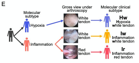
From: Tang et al., Nat Commun. (2024)
Clinical, blood and histological features
Age, sex, height, weight and course of the disease did not differ between subtypes. Ir had the highest proportion of hyperemia in the tendon and synovium. Ir also had the worst function:
- Function: lowest Oxford Shoulder Score, and poorer anteflexion, abduction, and external rotation than Hw. However, pain (VAS) did not differ.
- Imaging: highest Goutallier stage, indicating greater fat infiltration and consequently more structural damage.
- Blood: higher D-dimer, PT, and INR than Hw, which may suggest a hypercoagulable state or thrombosis. Hw had higher albumin, beta-hydroxybutyrate and glycyl-proline dipeptidyl aminopeptidase than Ir. Hw also had higher phosphorus than Iw, and Iw had higher indirect bilirubin than Ir.
- Histology: Ir had the highest vascularization (“veins”) score. Hw had higher GAG content.
Molecular mechanisms
Gene set variation analyses showed that neovascularization was downregulated in Hw and strongly upregulated in Ir. On the other hand, Hw upregulated hypoxia, glycolysis, and chondrocyte and cartilage development pathways and was termed the hypoxic atrophic subtype. Iw and Ir both upregulated inflammatory pathways, with Ir highest. Iw subtype was termed the intermediate inflammatory proliferative subtype, and Ir the high inflammatory proliferative subtype.
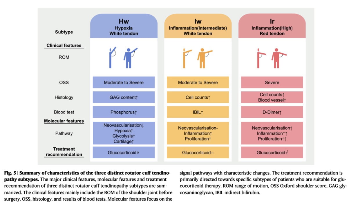
From: Tang et al., Nat Commun. (2024)
Animal models
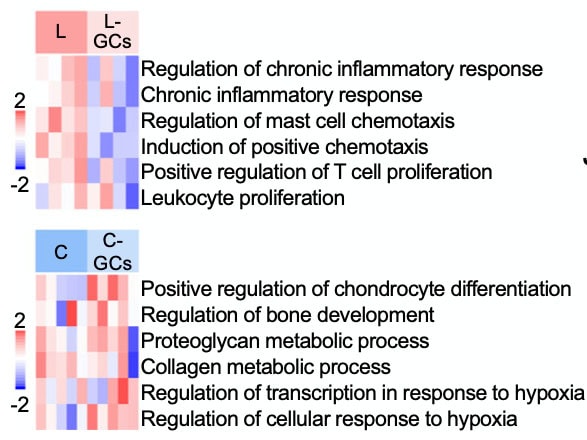
The levofloxacin rat model resembled human Ir, and the collagenase model resembled Hw. Glucocorticoids improved histology, collagen fibre diameter, treadmill performance, and inflammatory signatures only in the levofloxacin (Ir-like) model. They were harmful in the collagenase (Hw-like) model.
Clinical glucocorticoid data
After intraoperative glucocorticoid injection, Ir patients improved more than Hw patients in anteflexion and abduction at both 6 and 24 months. They also improved more on the “comb hair” OSS item at 6 months. This suggests glucocorticoids are effective mainly in the high-inflammatory (Ir) subtype.
Limitations
There were no matched healthy supraspinatus controls, and animal models only partly replicate human subtypes. Hw made up 53.7% of patients, which may help explain the conflicting glucocorticoid results in earlier literature.
Questions and Thoughts
The results suggest that patients classified in the Ir tendinopathy subgroup may experience greater improvement following glucocorticoid injection. At baseline, the Ir subtype showed the poorest function, with the lowest OSS and significantly reduced anteflexion, abduction, and external rotation compared with Hw. This clinical presentation might therefore point toward a highly inflammatory, hyperemic subtype, although it is unlikely to be specific enough to classify patients on its own.
At the structural level, glucocorticoid treatment showed promising effects in the animal model, including the downregulation of inflammation-related pathways. These findings suggest that glucocorticoids may be a potential treatment option for patients with an inflammatory tendinopathy profile. However, their clinical effectiveness remains uncertain. In this context, symptom-management strategies such as activity modification, temporary avoidance of provocative activities, and NSAIDs when appropriate may also be considered according to the patient’s individual presentation.
Interestingly, disease duration did not differ significantly between subgroups, suggesting that these molecular profiles may not simply reflect successive stages of tendinopathy. Pain intensity also did not differ significantly between the Ir, Iw, and Hw tendinopathy classification subtypes, highlighting that pain severity may not directly reflect the underlying tendon pathology.
A recently published systematic review further explores the mechanisms underlying tendinopathy-related pain. It describes contributions from mechanical load transduction, peripheral neuroimmune–vascular interactions, and the central nervous system (CNS). These interacting mechanisms may help explain the variability in patients’ pain experiences and support further investigation into whether more precise tendinopathy classification could help guide individualized treatment.
Talk nerdy to me
As described in the methods, the authors first collected baseline data for each patient: shoulder range of motion, OSS, MRI, blood tests, arthroscopic findings, and tendon RNA-seq, along with histology. NMF consensus clustering of the transcriptomes identified two molecular subtypes, H (hypoxia) and I (inflammation). Adding tendon colour under arthroscopy split Subtype I into Iw and Ir, giving three tendinopathy classifications: Hw, Iw and Ir.
We now examine the transcriptomic and histological characteristics of these subtypes, beginning with differential gene expression. The heatmap (Fig. F) shows the relative expression of the top 100 upregulated genes of each subtype, with red indicating higher expression and blue lower. Hw is characterized by hypoxia response genes, Iw by muscle contraction genes, and Ir by chemokine pathway genes.
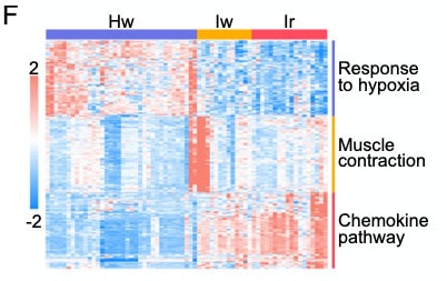
From: Tang et al., Nat Commun. (2024)
In Hw, HIF pathway genes (EGLN3, ALDOC, ENO1, ENO2) are upregulated, consistent with a shift toward anaerobic metabolism, while angiogenesis-related genes (FLT1, ANGPT1, ANGPT2) are downregulated, hence the term “hypoxia atrophy”. GAG biosynthesis genes are also upregulated, notably CHSY1 and CHPF (chondroitin synthesis). This fits the higher GAG content on Safranin O staining, which the authors interpret as possible chondrogenic remodelling.
In contrast, Iw and Ir show increased expression of chemokine genes (e.g., CCL11, XCL2, CCR4, CXCR1), migration genes (GNGT2, ITK, PRKCB, RAC2) and immune genes, including T-cell receptor components (CD3D, CD3E, CD247, CD4) and signalling molecules (LCK, LCP2, BTK). This is consistent with an immune response and immune cell infiltration, most pronounced in Ir, which also shows upregulated neovascularisation.
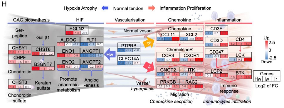
From: Tang et al., Nat Commun. (2024)
Finally, glucocorticoid treatment reduced inflammatory signatures in the rat levofloxacin model, the closest match to human Ir. The heatmap shows lower scores for pro-inflammatory pathways (chronic inflammatory response, mast cell chemotaxis, T-cell and leukocyte proliferation) after treatment. No comparable reduction is seen in the collagenase model, which resembles Hw. L: levofloxacin model; L-GCs: levofloxacin model with glucocorticoids; C: collagenase model; C-GCs: collagenase model with glucocorticoids.
From: Tang et al., Nat Commun. (2024)
Take-home messages
- Tendinopathy is not one disease. Transcriptomic profiling plus tendon color under arthroscopy identified three rotator cuff subtypes with distinct biology: Hw (hypoxic atrophic), Iw (intermediate inflammatory proliferative) and Ir (high inflammatory proliferative).
- Ir is the most severe clinical phenotype. It combines the highest inflammation and neovascularisation with the worst shoulder function, the highest Goutallier stage, and a possible hypercoagulable blood profile. Pain intensity and disease duration did not differ between tendinopathy classifications, so neither is a reliable marker of the underlying tendon pathology.
- Glucocorticoids may help only some patients. In rats, glucocorticoids reduced inflammatory signatures and improved tendon structure only in the Ir-like model, and were harmful in the Hw-like model. In patients, Ir improved more than Hw after intraoperative injection. Because all patients received the injection and there was no control group, these findings are suggestive rather than proof of subtype-specific efficacy.
- Clinical implication. A one-size-fits-all approach may explain the conflicting glucocorticoid results in the literature, as Hw made up over half of the cohort. Subtype-guided treatment is a promising direction, but prospective validation studies and practical, non-invasive ways to identify tendinopathy classification subtypes are still needed.
Reference

