

Research
Ankle/Foot
July 24, 2026
From Generic to Specific: How to Prescribe Patient-Specific Achilles Tendinopathy Rehabilitation?
This biomechanical modelling study gives insight into how patient-specific characteristics influence Achilles tendon strains in those with midportion Achilles tendinopathy
Based on the factors influencing tendon strain, exercises can be ranked, and an individual exercise plan can be established using this information
Although this was not a clinical study, we try to break it down into practical implications
Introduction
Although we know that Achilles tendons require proper loading and that several rehabilitation models have been developed, many patients experience poor results after Achilles tendinopathy rehabilitation and develop more chronic tendinopathy. In practice, loads are prescribed and progressed according to tendon capacity, tissue irritability, and symptom response. We know that in general, bilateral exercises are easier than unilateral exercises, and that hopping exercises are more demanding than non-hopping ones.
While we already know much about loading factors, the factors we consider in practice are mostly based on external exercise doses, neglecting the influence of the internal patient-specific tendon properties. Most rehabilitation decisions are based on the external exercise dose like the exercise selected, the number of repetitions, the external resistance, the speed, and the range of motion. Much less clear is how that prescribed task translates into the internal mechanical dose experienced by the tendon.
This paper gives a biomechanical explanation of why an exercise can be beneficial for one patient, while the same exercise in another patient can exert little to no effect. We try to break down this biomechanical study into clinical examples and takeaways.
Methods
Funaro et al. conducted a cross-sectional, subject-specific finite-element modelling study to estimate internal Achilles tendon strain during six common rehabilitation exercises in people with mid-portion Achilles tendinopathy.
Participants with a history of intermittent episodes of Achilles tendinopathy pain for more than 6 consecutive weeks within the past 5 years were included if they had at least one episode of tendon pain exacerbation and remission within that period. Furthermore, they had to have a palpable and painful focal thickening of the Achilles tendon in the midportion, which was supported by sonographic evidence of Achilles tendinopathy.
Candidates were excluded from the study when they had previous Achilles tendon surgery or tears, systemic diseases affecting collagen tissue or insertional Achilles tendinopathy. Also, those with calcaneal spurs, plantar fasciitis or other conditions of the foot and ankle were excluded.
All included participants completed the Victorian Institute of Sport Assessment-Achilles questionnaire (VISA-A), to get insight into their pain and function.
Participants first performed maximal plantarflexion contractions on an isokinetic dynamometer. Three-dimensional freehand ultrasound was then used to assess the Achilles tendon at rest and during contractions at 30% and 60% of maximum effort. From these measurements, the researchers estimated tendon length, elongation, cross-sectional area, force, and stiffness for each participant.
During a second laboratory session, participants performed five repetitions of six common rehabilitation exercises in random order: bilateral heel rise, bilateral heel drop, unilateral heel drop with the knee extended, walking, unilateral heel drop with the knee flexed and bilateral hopping. Motion capture, force-plate data and surface EMG were combined with musculoskeletal modelling to estimate the forces produced by the soleus and both gastrocnemius muscles during each exercise.
These measurements were used to create a personalised computer model of each participant’s Achilles tendon. The main model included individual tendon shape, stiffness and muscle forces, allowing the researchers to estimate strain within the midportion of the Achilles tendon and rank the exercises from lower to higher strain.
Two additional models were created to explore what explained the differences between the participants. One retained individual tendon stiffness but used average muscle forces, while the other retained individual muscle forces but used average tendon stiffness. This allowed the authors to compare whether predicted tendon strain was influenced more by muscle-force production or by tendon properties. Although the outer tendon shape was personalised, the internal subtendon arrangement and twist were based on a generic anatomical model rather than being measured individually.
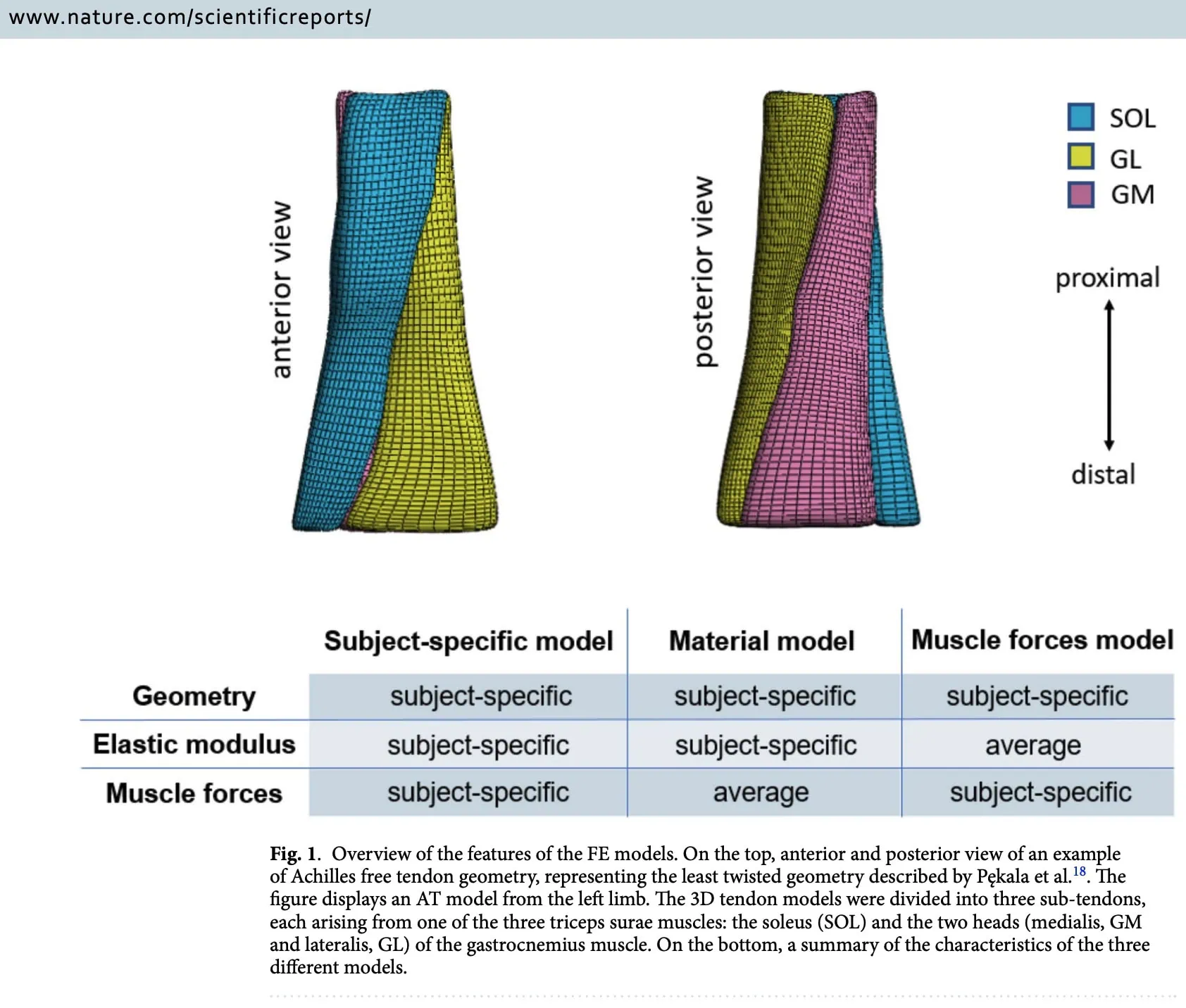
The primary outcome of this study was the average maximum strain in the midportion of the Achilles tendon for each of the six exercises in these participants with Achilles tendinopathy. Then, the authors also examined the peak local strain and its location, as well as the variation in strain among participants. A strain range of 5-7% was used as the optimal strain required for tendon adaptation, based on prior research evidence.
Importantly, all measurements were obtained prior to starting any rehabilitation plan. In this way, this study reflects the Achilles tendon properties and response to loading at the painful tendinopathy stage before the introduction of a rehabilitation plan.
Results
Twenty-one participants with midportion Achilles tendinopathy were recruited, of whom 17 were males, and 4 were females. Their mean age was 49 years (+/-13 years), they were, on average, 178 cm (+/-8 cm) tall and weighed 75 kg (+/- 12kg). Their mean VISA-A score was 73 (+/- 19).
Starting from the lowest average midportion Achilles tendon strain to the highest, the following ranking was revealed:
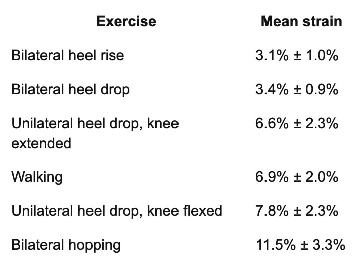
All exercises were statistically different from one another, except walking and unilateral heel drop with the knee extended, which produced similar strains.
Based on this result, the group averages show that unilateral heel drop with an extended knee and walking provide the strain necessary to promote tendon adaptation.
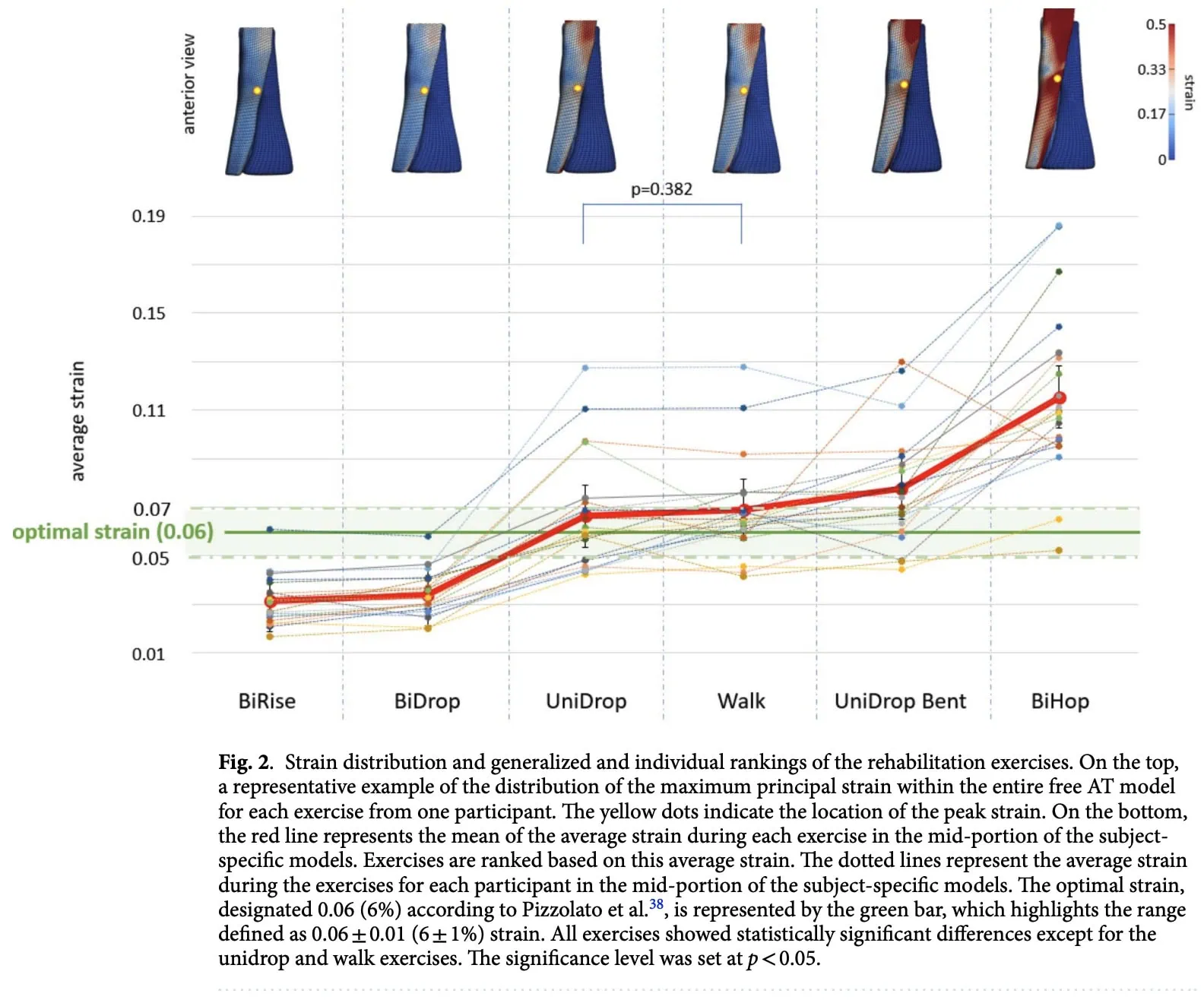
An interesting finding arose when Achilles tendon strain at the individual patient-specific level was compared with the optimal strain necessary for tendon adaptation based on the 5-7% necessary strain. Some reached the 5–7% range during easier exercises such as bilateral heel rises, whilst others only reached it during more demanding tasks such as a flexed-knee heel drop or hopping. The order of exercises from low to high strain also differed between some participants.
The number of participants who reached the proposed 5-7% strain range during each exercise was:
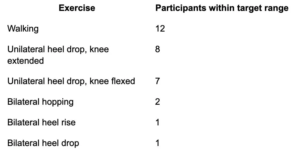
It appeared that some required more demanding exercises to get to the optimal Achilles tendon strain.
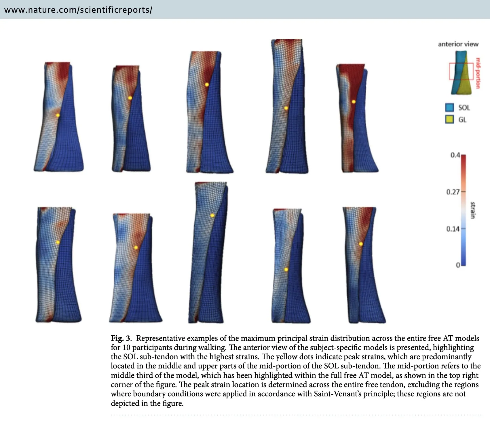
The computer models showed very high strain in a few small areas of the tendon, especially during flexed-knee heel drops and hopping. These numbers should not be read as if the entire tendon stretched by that amount. They represent tiny local “hot spots” within the model and are sensitive to assumptions about tendon shape, subtendon borders, and how the model was built. These local peaks were usually found in the middle or upper-middle part of the tendon, near the soleus subtendon. The exact location differed between patients. Clinically, this suggests that tendon loading is not distributed evenly, but the peak values themselves are not ready to guide exercise prescription.
The researchers also asked what explained the differences in strain between patients. They found that keeping each person’s own calf-muscle forces produced results much closer to the fully personalised model than keeping only each person’s tendon stiffness. This does not mean tendon stiffness is unimportant. It means that, in these models, individual force production had a greater effect on predicted strain.
Questions and thoughts
Importantly, this was a computational modelling study. It did not test whether prescribing exercises according to estimated tendon strain improves pain, function, recovery time or return to sport. Yet, the information derived from this study can help us in our rehabilitation process.
We already know a lot about exercise dosing, as we already manipulate external loads, repetitions, sets, tempo, range of motion, frequency, task complexity, and we already know that rehabilitation should be individualized. So what is genuinely new here? The study gives us new understanding of the difference between external and internal tendon loads. We prescribe our patients with tasks that give an external load to the Achilles tendon, and the tendon experiences an internal mechanical stimulus. These are related but not identical to one another. We can divide the loads into three levels:
- Prescribed dose: What the physiotherapist selects
- Exercise
- External resistance
- Sets and repetitions
- Tempo
- Frequency
- Performed dose: What the patient actually produces
- Range of motion
- Force
- Movement speed
- Effort
- Compensations
- Fatigue
- Internal tendon dose: What the tendon experiences
- Strain magnitude
- Strain distribution
- Loading rate
- Duration
- Cumulative mechanical exposure
Clinical practice mainly controls the first level and observes parts of the second. This study attempted to estimate the third. As such, this study does not overturn common rehabilitation principles. Instead, it warns against assuming that a standard progression translates into a standard biological dose.
The most important challenge for us is how to determine which load in which patient yields the optimal strain necessary for tendon adaptation. This study was a biomechanical analysis and provided important information regarding the stimulus needed for tendon adaptation in tendinopathic Achilles tendons, but it couldn’t provide a “recipe” for the individual patient. So how can we use this information in practice, and how should it alter our practice?
The generic hierarchy remains clinically useful: starting with bilateral work, progressing to unilateral work, to flexed-knee loading, to hopping. But it should be used as a starting point, rather than a fixed rehabilitation framework. Clinically, we can not determine the “optimal” load for each patient specifically, but we can try to estimate whether the training dose is insufficient, productive, or excessive.
Let’s break it down into some clinical examples:
Patient A: low-capacity, irritable tendon
Imagine a patient with:
- pain during daily walking;
- poor bilateral calf-raise capacity;
- marked fatigue;
- a strong next-day symptom response.
For this person, a bilateral heel raise may already be a meaningful tendon stimulus.
The study showed that although bilateral exercises produced low strain on average, some participants reached the proposed target range during them. Do not dismiss bilateral work as “too easy” simply because it appears early in a protocol. Instead, begin with an exercise the patient can tolerate, for example:
- supported bilateral calf raises;
- seated plantarflexion;
- reduced range;
- slower tempo;
- lower volume.
Progress when the patient demonstrates improved capacity and recovery, not merely because a timetable says that unilateral exercise should begin.
Patient B: strong patient who is underloaded
Imagine an athletic patient who:
- performs bilateral and unilateral calf raises easily;
- shows little fatigue;
- reports low effort;
- has minimal response to the programme;
- remains below sport demands.
For this patient, standard calf raises may not produce enough force to challenge the tendon meaningfully. Check whether the exercise is truly demanding:
- Is sufficient external load being used?
- Is full heel-rise height maintained?
- Does performance decline across the set?
- Is the final repetition close to fatigue?
- Is the patient shifting weight to the other leg?
- Are both straight-knee and flexed-knee plantarflexion being trained?
Progress through:
- increased external resistance;
- unilateral loading;
- greater range;
- flexed-knee loading;
- faster loading;
- hopping;
- sport-specific energy-storage tasks.
Completing the exercise does not prove that the tendon received an adequate stimulus.
Patient C: unexpectedly strong response to a “reasonable” exercise
Imagine a patient who tolerates straight-knee unilateral heel raises but flares after:
- flexed-knee heel drops;
- hopping;
- a small increase in speed or volume.
The paper estimated that flexed-knee unilateral heel drops generally produced greater strain than straight-knee unilateral heel drops, while hopping produced the highest strain. But it also showed considerable individual variation. So, do not interpret the flare automatically as fear, poor adherence, or treatment failure. The exercise may represent a larger internal mechanical step for that patient than expected.
Modify:
- knee position;
- external load;
- range;
- movement speed;
- repetition count;
- number of sets;
- weekly frequency.
For example, replace a demanding unilateral flexed-knee heel drop with a lighter seated calf raise and build capacity before returning to the original task. A small-looking exercise modification may cause a large change in tendon loading.
Talk nerdy to me
A very important caveat is that this study was not assessing how the participants progressed over time; rather, they were assessed at one particular point in time, before they even started rehabilitation for their Achilles tendinopathy. We can thus not assume that using this information will produce better rehabilitation outcomes.
An important limitation of this study is that it used a proposed optimal strain range, based on healthy individuals and animal and in-vitro studies. But a pathological tendon may require a different loading range. The dose may also vary according to the irritability and capacity of the tendon, the chronicity of the complaint, the loading history, and the rehabilitation phase.
Another important aspect of the current study that warrants careful consideration is that the study assessed the strain once, during some repetitions, but it did not assess the exercises throughout a specified amount of time. So, for example, walking fell within the proposed target range, but the study didn’t assess the speed of walking, the step count (cadence), the duration, the inclination, et cetera. So the adaptation of the Achilles tendon is not only dependent on the strain magnitude, but is also time- and rate-dependent.
The study focused on tensile loads and strains, but there is always a proportion of compressive load to the tendon as well. Furthermore, the friction contact between sub-tendons was dismissed, which simplified the analyses, but can be a limitation.
Take-home messages
This paper does not provide a new Achilles rehabilitation protocol. It provides a more precise way to think about exercise dose. The average exercise hierarchy was largely predictable: bilateral calf work produced the lowest estimated tendon strain, unilateral loading produced more, flexed-knee unilateral loading produced still more, and hopping produced the highest strain. The most important finding was that individual patients did not always follow that average pattern.
The same exercise could represent a relatively low, moderate or high internal tendon load depending on the patient’s:
- muscle-force production;
- tendon geometry;
- cross-sectional area;
- material properties.
The practical change is therefore not to abandon existing rehabilitation principles, but to become less confident that an exercise label defines the load. A bilateral exercise may be too easy for some, but just enough for others. As physiotherapists, we should aim to prescribe patient-specific exercises for Achilles tendinopathy rehabilitation, not just generic protocol-based exercises. By manipulating the external loads, which we already do, and being aware of the patient-specific internal loads, we should be able to individualize our exercise prescription according to the patient in front of us, rather than just following a generic progression.
The major limitation is that the study assumes approximately 5–7% strain is therapeutically optimal, although this has not been demonstrated in people with Achilles tendinopathy. The study shows which exercises were predicted to reach that range. It does not show that reaching it improves outcomes.
Reference
WHAT TO LOOK FOR TO PREVENT HAMSTRING, CALF & QUADRICEPS INJURIES
Whether you’re working with high-level or amateur athletes you don’t want to miss these risk factors which could expose them to higher risk of injury. This webinar will enable you to spot those risk factors to work on them during rehab!