

Learn
Cuboid Syndrome | Chronic Lateral Ankle Pain Assessment & Treatment
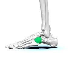
Ankle sprains account for a big portion of lower extremity injuries with up to 40% of cases having residual symptoms. There’s a hypothesis that the cuboid may be responsible for those cases in which lateral ankle pain persists. It appears that the condition has a higher prevalence in professional ballet dancers. The hypothesis for cuboid syndrome is based on history clusters of signs and symptoms, differential diagnosis, clinical expertise, and of course mechanism of injury. It’s assumed that during a severe initial inversion trauma, torsion between the cuboid and navicular bone and cuneiform as well as the calcaneus leaves the cuboid in a relatively supinated position.
This disposition remains painful specifically over the calcaneocuboid joint while tenderness over lateral ankle ligaments subsides. Patients display antalgic gait with an increase in pain during the push-off phase and if pain allows for mobility testing, joint play is absent. A technique that’s described in the literature is the reposition manipulation into relative pronation.
It’s reported that following the manipulation patients were pain-free the following day and could return to their activities. For the technique, the patient lies supine with the leg extended and the foot hanging over the edge of the bench.
You’re going to position yourself medially to the foot and place the hypothenar of your contralateral hand on the lateral and plantar aspect of the cuboid just proximal to the base of the fifth metatarsal. The hypothenar on the other hand is placed dorsally and medially on the cuboid which is lateral and proximal to the line between the third and the fourth digit.
In this position, the forearms form a straight line and after you fold your hands together ask the patient to make active maximal dorsiflexion which pretensions the cuboid submaximal eversion. Build up compression through your elbows as well as increase the pronation pre-tension direction.
Ask the patient to relax the foot and then perform a pronation swing with both elbows combined with a compression impulse of both hypothenars.
As previously mentioned differential diagnosis is necessary and the first step of assessing ankle sprains should be to exclude a fracture. So make sure to read our wiki on the Ottawa ankle rules next.
Get the 21 most useful orthopedic tests
Download our free e-book with the 21 most useful orthopedic tests per body region, to help you reach the right diagnosis.

Like what you’re learning?
Get the full Physiotutors Assessment Book
- 600+ pages on orthopedic assessment
- Video demonstrations and direct links to the PubMed studies
- Statistical values for every special test, from the latest research
- Free lifetime updates
- Available in English, German, French, Spanish, Italian, Portuguese and Turkish