


Research
Ankle/Foot
September 16, 2026
Chopart Injuries: A Key Differential Diagnosis Not to Miss in Physiotherapy Practice - Highlights on Diagnosis and Management
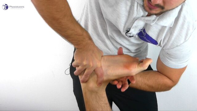
X-rays often underestimate or miss Chopart injuries entirely. A CT scan is indicated when suspicion is high
Plantar ecchymosis is a red flag, not a rule-out: it's a strong indicator of significant midfoot injury, but its absence doesn't exclude one
ORIF looks better, but evidence is inconclusive: open reduction and internal fixation tends to outperform closed reduction
Introduction
Ankle and foot injuries are commonly encountered in physiotherapy practice. The Ottawa Ankle Rules provide a useful clinical decision tool to identify patients who may require radiographic assessment following an acute ankle or foot injury. However, some midfoot injuries can remain challenging to identify during the initial assessment. In a previous Physiotutors article, we discussed Lisfranc injuries, which can be missed during initial medical encounters and on plain radiographs, potentially contributing to persistent symptoms and functional impairments.
This article focuses on Chopart injuries, another potentially serious midfoot injury that is considerably less common but can have important implications for diagnosis, medical management, and rehabilitation. Given their potential to be overlooked and the limited evidence available, recognizing the clinical presentation and understanding current management strategies are particularly relevant for physiotherapists.
This literature review provides an evidence-based overview of the diagnosis and management of Chopart injuries, with particular attention to considerations relevant to physiotherapy practice.
Methods
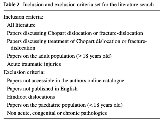
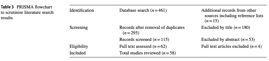
To conduct this literature review, medical databases were systematically searched for Chopart injuries, following a PRISMA-informed approach. Given the scarcity of available evidence, no predefined eligibility criteria were applied during the initial literature search. Following this initial identification phase, the retrieved literature was screened, and inclusion and exclusion criteria were subsequently applied, as detailed in Table 2. This process resulted in the inclusion of 58 studies.
Results
Among the 58 included studies, 36 were case reports, 4 were cohort studies, and 4 were case series. The remaining 14 studies addressed the epidemiology, diagnosis, treatment, and outcomes of Chopart dislocations and fracture-dislocations.
Anatomy
As the original study provides only limited anatomical information in its introduction, we will first review the relevant anatomy of the Chopart joint.
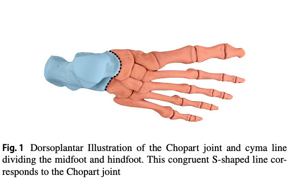
The midtarsal joint, also known as the Chopart joint, was named after the French surgeon François Chopart, who described an amputation technique through the articulation between the hindfoot and midfoot. The Chopart joint consists of two articulations: the talonavicular joint (TNJ) and the calcaneocuboid joint (CCJ). Together, these articulations form the characteristic S-shaped “cyma line” that defines the Chopart joint, as illustrated in Figure 1.
The TNJ is supported superiorly by the dorsal talonavicular ligament and the medial component of the bifurcate ligament, while the spring ligament provides inferior support.
Similarly, the CCJ is supported superiorly by the dorsal calcaneocuboid ligament and the lateral component of the bifurcate ligament. Inferiorly, the short plantar ligament contributes to plantar stability of the joint.
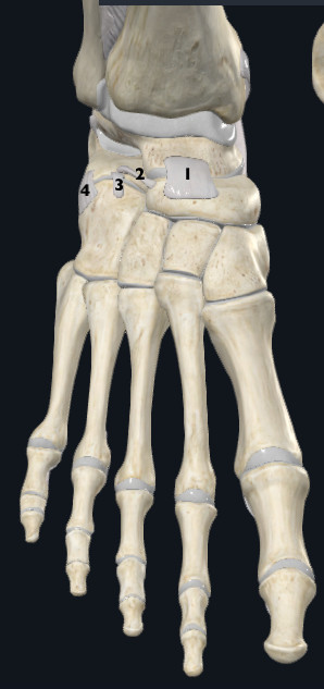
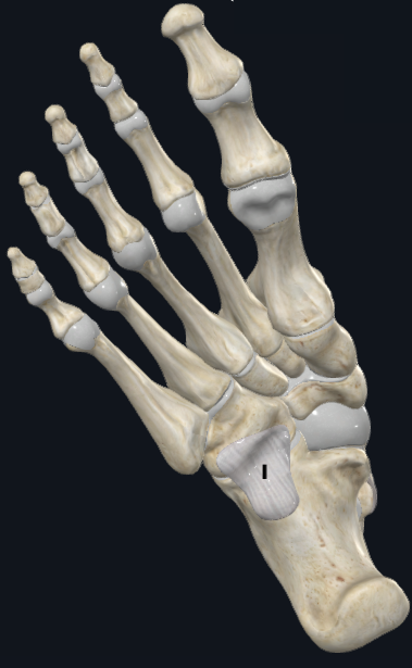
The TNJ contributes to pronation and supination of the tarsus, as it forms part of the talocalcaneonavicular joint. The CCJ, on the other hand, allows approximately 25° of rotation associated with hindfoot inversion and eversion. Together, the two articulations allow the forefoot to remain inverted or everted relative to the hindfoot, effectively locking the TNJ and CCJ and providing midfoot stability during the push-off phase of gait.
Chopart injuries can be broadly classified into four groups according to the structures involved: ligamentous injuries with or without dislocation, and fractures with or without dislocation, affecting either or both the CCJ and TNJ. A pure dislocation refers to dislocation of the navicular and/or cuboid without an associated fracture. A fracture-dislocation involves a fracture of the navicular and/or cuboid, with associated fractures involving one or more of the talus, calcaneus, navicular, or cuboid.
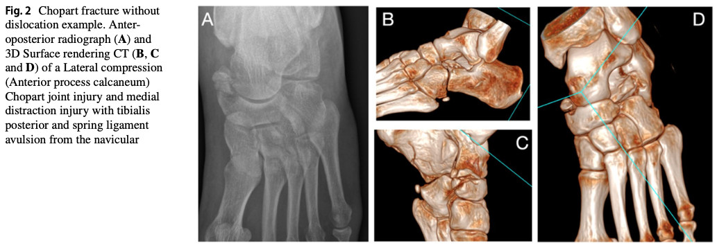
Fractures must occur at one or more of the following locations: the talus, calcaneus, cuboid, or navicular bone, including avulsion fractures, as illustrated in Fig. 2.
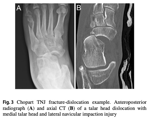
Chopart injuries could be classified according to the direction and the magnitude of force. The following table describes the types and descriptions of injuries. To note, swivel dislocation refers to TNJ and/or CCJ dislocation resulting from a medial or lateral deforming force. In a swivel-type Chopart dislocation, the talocalcaneal ligament remains intact and acts as a pivot around which the calcaneus and the distal foot rotate relative to the talus.
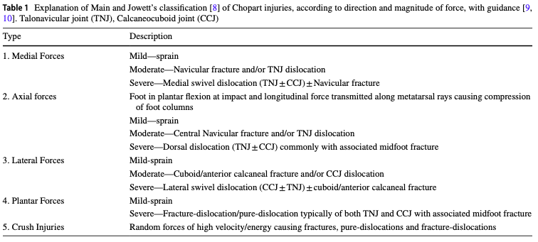
Chopart injury etiology
Chopart injuries are rare, with one epidemiological study reporting an incidence of 2.2 per 100,000 people per year. Motor vehicle accidents are reported as the primary etiology. In a study of 110 patients with Chopart injuries, 25% presented with a pure dislocation, 55% with a fracture-dislocation, and 20% with a combined Lisfranc and Chopart fracture-dislocation. However, these findings should be interpreted with caution, as another study of 128 patients reported a substantially lower proportion of pure dislocations (3.7%).
Diagnosis
Only a few of the included studies reported their diagnostic methods. In the two studies that did, both X-ray and CT scans were performed. Interestingly, the diagnosis of Chopart injuries is frequently delayed, and initial X-ray interpretation may underestimate the extent and severity of the injury, with CT providing a more comprehensive assessment.
Management
Among the available literature, management strategies varied according to injury type.
Fracture-dislocations: 83% of the 60 reported injuries were managed surgically. Open reduction and internal fixation (ORIF) was performed, with or without the use of external fixation.
Pure dislocations: Of the 28 reported cases, 19 were managed with closed reduction, while 6 patients required additional procedures, including internal and/or external fixation.
Regarding complications, compartment syndrome was occasionally reported following midfoot and Chopart injuries, as well as Chopart dislocations, and required fasciotomy when present.
Interestingly, primary amputation was reported in approximately 5% of fracture-dislocations and 25% of combined Lisfranc and Chopart fracture-dislocations. In a cohort of 128 patients, 7 required arthrodesis of the Chopart joint, while approximately 5% underwent delayed fusion of the Chopart joint.
Outcomes
The AOFAS scale was used to assess outcomes. AOFAS measures pain, function, and foot alignment. The total score is rated out of 100, with a score of 100/100 indicating a perfectly healthy ankle and foot.
Regarding injury types, here are the outcomes reported on the AOFAS:
- Isolated Chopart fractures: 81/100 (1 study, n=28)
- Chopart fracture-dislocations: 70.3/100 (3 studies, n=54)
- Chopart pure dislocations: 79/100 (1 study, n=14)
- Combined Chopart-Lisfranc fracture-dislocation: 58/100 (2 studies, n= 54)
Across all groups, better outcomes were observed in patients who underwent ORIF. At 9-year follow-up, Chopart fracture-dislocations were associated with more frequent radiographic abnormalities and post-traumatic arthritic changes than isolated Chopart fractures. Structural abnormalities, particularly incorrect medial and lateral column lengths, were strongly associated with poorer gait quality, whereas post-traumatic arthritis appeared to have little influence on gait.
Several negative prognostic factors were identified, including motor vehicle accidents, work-related incidents, open injuries, polytrauma, associated fractures, and delayed surgery (>1 day after injury). ORIF performed as the initial treatment was associated with better outcomes than internal fixation following closed reduction.
Regarding case reports, the findings suggest that CT scans are often required for adequate assessment of Chopart injuries, while closed reduction has a substantial failure rate. Long-term complications, particularly pain and malunion, are also common despite surgical treatment.
Question and thoughts
Clinical examination should aim to identify plantar ecchymosis, which has been described in the literature as a pathognomonic sign of significant midfoot injury. Plantar ligament disruption can result in hematoma formation and subsequent plantar ecchymosis. However, a pathognomonic sign is not necessarily sensitive; therefore, the absence of plantar ecchymosis does not exclude a significant midfoot injury.
Imaging is also critical, as Chopart injuries are frequently missed at initial presentation. Plain radiographs may underestimate the extent and severity of the injury or fail to identify the injury altogether. CT is therefore recommended when a Chopart injury is suspected to provide a more comprehensive assessment of the osseous and articular involvement.
Regarding management, as discussed in the results section, the available evidence supports ORIF as the preferred approach when surgical treatment is indicated. Closed reduction alone appears to be associated with poorer long-term functional outcomes and should therefore be approached with caution. Evidence regarding physiotherapy management remains limited. Restoration and maintenance of appropriate medial and lateral column alignment appear to be particularly important for optimizing long-term gait function.
Talk nerdy to me
As highlighted by the authors, the literature on Chopart injuries remains particularly scarce and heterogeneous. The evidence supporting surgical outcomes is largely based on a limited number of cohort studies, with some of the key evidence dating back several decades.
The study most frequently cited regarding the superiority of ORIF was published in 2004 and was based on a retrospective cohort. The patients included in this study were therefore treated according to surgical practices from several decades ago. Although the second major study was published in 2023, it was a prospective cohort with a mean follow-up of 10.2 years, meaning that a substantial proportion of its findings also reflects treatment practices from the preceding decade.
The other studies contributing to the outcomes section were published between 1997 and 2014 and were generally less influential in supporting the superiority of ORIF. Overall, the evidence supporting current surgical recommendations therefore remains based on a relatively small and heterogeneous body of literature, with much of the evidence originating from older cohorts. Consequently, the magnitude of the reported benefit of ORIF should be interpreted cautiously, as contemporary surgical techniques and peri-operative management may differ from those used in the original cohorts.
Interpretation of the case reports is also limited. Although several of these reports are more recent, the evidence is based on very small samples and highly heterogeneous outcome reporting. In the fracture-dislocation case reports, for example, only 7 of 24 cases reported an AOFAS score, while many other reports relied primarily on descriptive clinical outcomes such as pain, stiffness, functional limitations, or return to activity. This limits the ability to make reliable comparisons between treatment strategies or to draw conclusions regarding long-term functional outcomes.
Take-home messages
- Think beyond the ankle: Chopart injuries are rare but potentially serious midfoot injuries that can be missed during the initial assessment.
- A normal X-ray does not rule it out: Clinical suspicion should remain high, particularly after high-energy trauma. CT may be required to fully assess the extent of the injury.
- Look for plantar ecchymosis: Although described as a highly suggestive sign of significant midfoot injury, its absence does not exclude a Chopart injury.
- Alignment matters: Restoration and maintenance of the medial and lateral foot columns appear to be important for preserving foot function and gait quality.
- ORIF appears to provide better outcomes: Available evidence generally favours open reduction and internal fixation over closed reduction followed by fixation, although the evidence base is limited and largely derived from older, heterogeneous studies.
Physiotherapy evidence is scarce: Rehabilitation should therefore be guided by the surgical procedure, tissue healing, restoration of mobility and strength, and progressive return to weight-bearing and functional activities. Interpret the evidence cautiously: Chopart injuries are uncommon, and current recommendations are largely based on retrospective cohorts and case reports rather than high-quality comparative studies.
Reference

