

Research
Ankle/Foot
August 25, 2026
Lisfranc Injury Diagnosis and Management: Key Considerations for Physiotherapists
This narrative review provides an overview of the clinical assessment, imaging, management, and rehabilitation of Lisfranc injuries
Lisfranc injuries are frequently missed or misdiagnosed, potentially resulting in delayed treatment, persistent pain, functional deficits, and poorer outcomes.
Early recognition and appropriate imaging are essential, particularly when a rotational injury occurs on a plantarflexed foot, as maintaining anatomic alignment is strongly associated with better outcomes
Introduction
Ankle sprains are among the most common conditions encountered in physiotherapy practice. Due to the anatomy of the ankle complex, the lateral ankle ligaments are particularly vulnerable to injury, with inversion on a plantarflexed foot being the most common mechanism. However, this mechanism can also place substantial stress on the midfoot, particularly the dorsomedial tarsometatarsal region, potentially involving the Lisfranc ligamentous complex, a key stabilizer of the midfoot.
Lisfranc injuries require specific management, often including a period of non-weight-bearing, yet they are frequently missed or misdiagnosed. Although traditionally considered rare, recent evidence suggests that their incidence may be higher than previously thought, particularly in athletic populations.
This review aims to provide physiotherapists with practical clinical knowledge to recognize, assess, and appropriately refer patients with suspected Lisfranc injury.
Methods
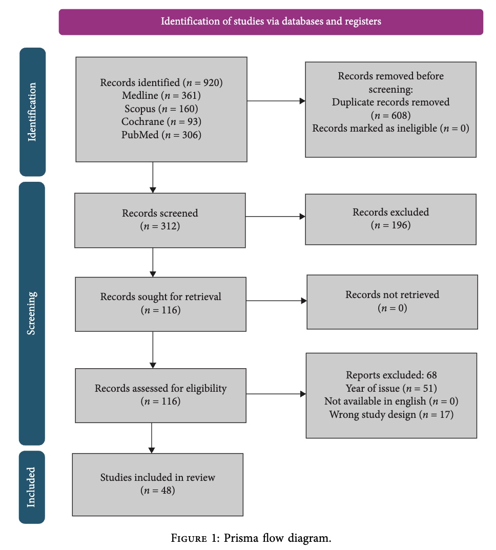
This study is a retrospective narrative review. Medical databases were systematically searched following the PRISMA guidelines and search strategy. Comparative and observational studies were included without age restrictions, while case reports, animal studies, and expert opinions were excluded. A total of 48 studies were included in the final review.
Results
Anatomy
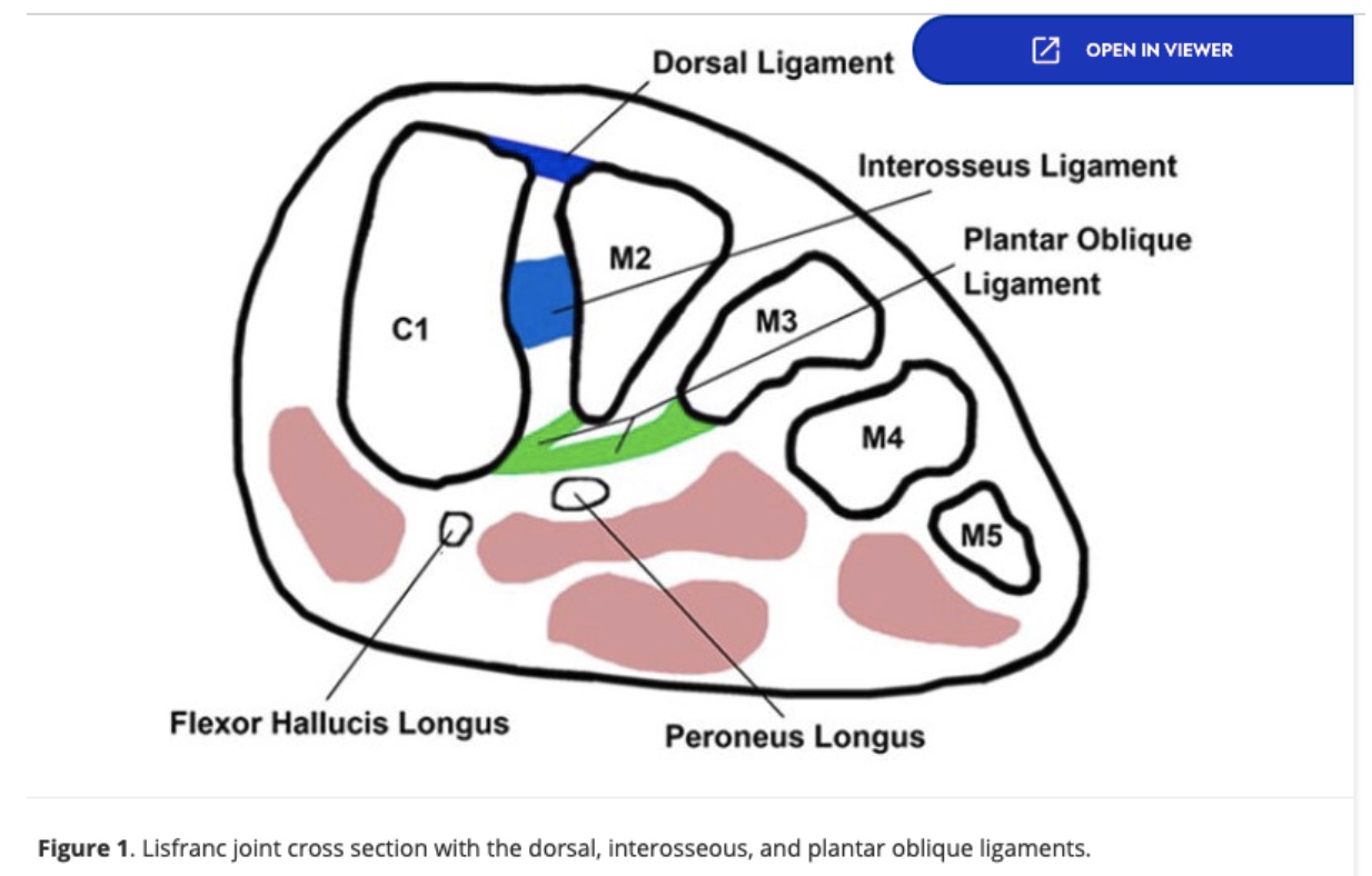
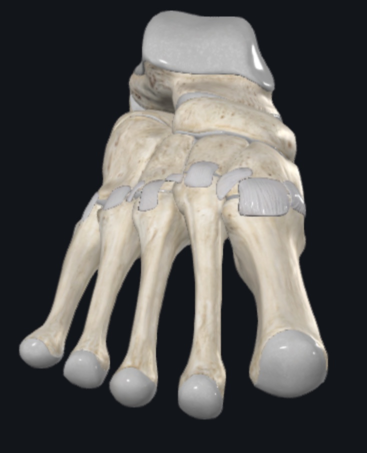
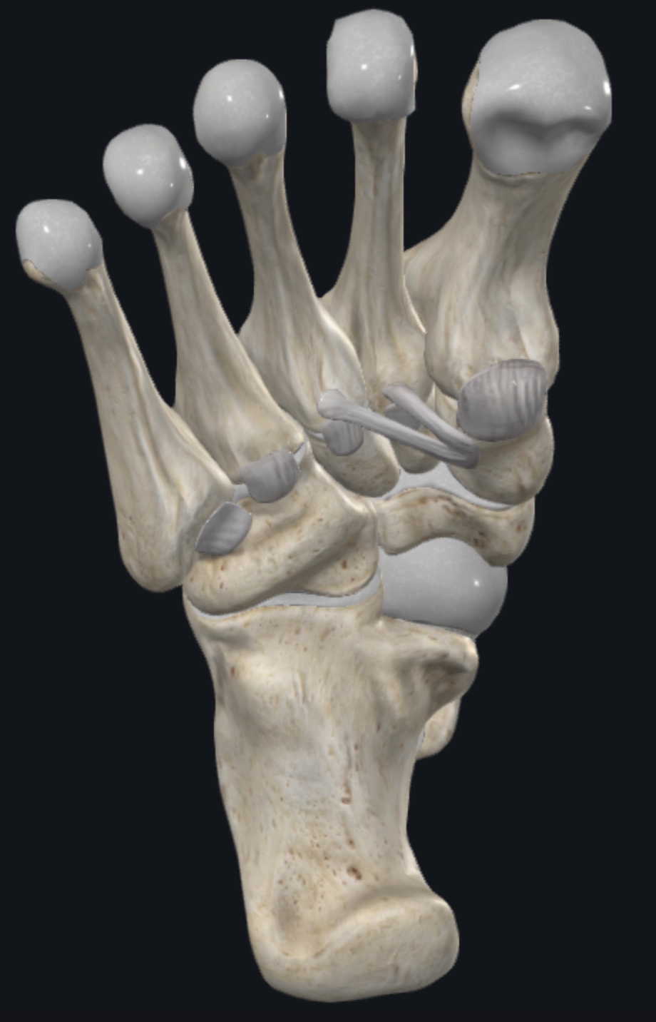
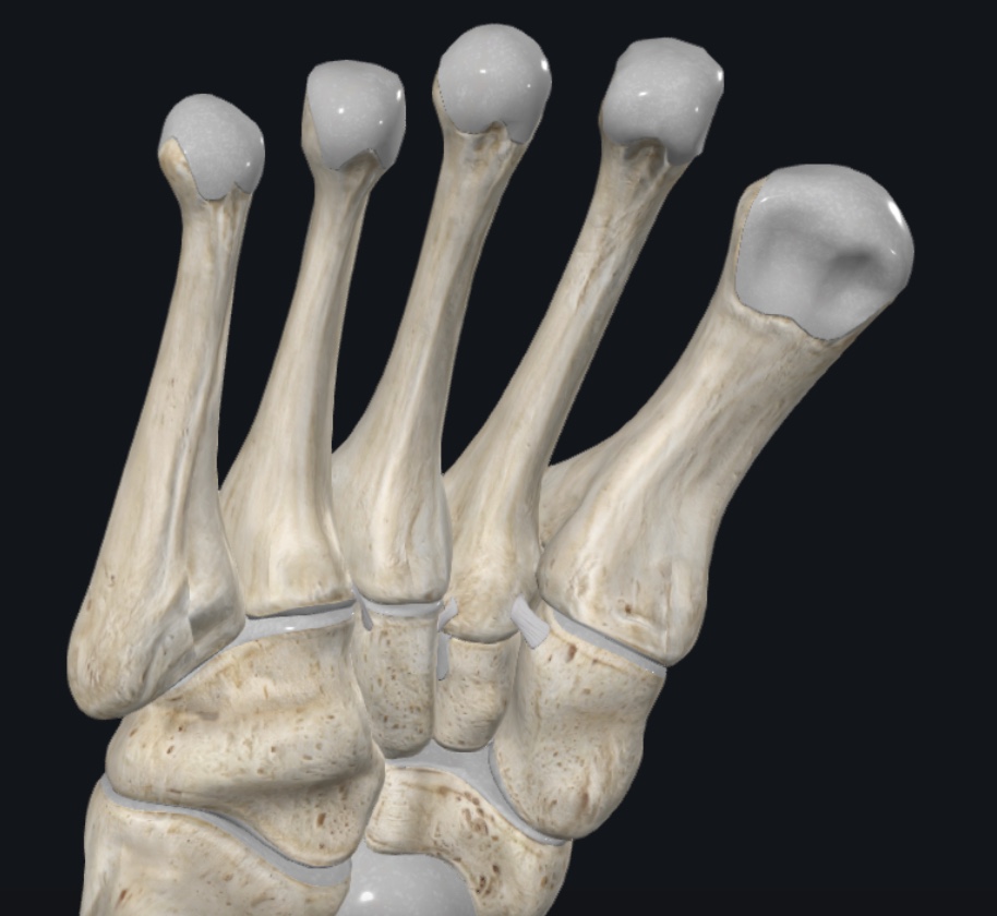
The Lisfranc joint complex comprises the articulations between the three cuneiforms and cuboid and the bases of the five metatarsals. The alignment of these bones provides structural stability to the medial and lateral foot arches and is reinforced by the Lisfranc ligament complex, which consists of the dorsal (DLL), interosseous (ILL), and plantar (PLL) ligaments.
The dorsal and plantar ligaments originate from the cuneiforms and attach primarily to the bases of the metatarsals, while the interosseous Lisfranc ligament originates from the medial aspect of the medial cuneiform and inserts onto the base of the second metatarsal. A plantar extension also contributes to the attachment toward the third metatarsal.
The Lisfranc complex is particularly important for midfoot stability because there is no direct interosseous ligament between the first and second metatarsals. Consequently, the DLL, ILL, and PLL provide much of the ligamentous stability of the tarsometatarsal region.
Incidence, etiology and biomechanics
Recently published data suggest that nearly 30% of Lisfranc injury diagnoses are initially missed, particularly following low-energy mechanisms or in polytrauma patients. In collegiate football players, Lisfranc injuries may account for up to 20% of foot injuries.
The typical mechanism involves rotational stress applied to a plantarflexed foot, which can result in disruption of the dorsal ligaments and/or fracture at the metatarsal base. Other common mechanisms include forefoot twisting, crush injuries, and high-energy trauma.
Biomechanical studies suggest that the interosseous Lisfranc ligament (ILL) is the stiffest component of the Lisfranc ligament complex. Disruption of this ligament can therefore substantially compromise midfoot stability and may require surgical fixation when adequate stability cannot be maintained conservatively.
Diagnosis, imaging and physical examination
Following trauma, patients typically present with midfoot pain, swelling, and difficulty with weight-bearing. Plantar ecchymosis is particularly suggestive of a Lisfranc injury diagnosis.
Clinical examination should include the piano-key test, first metatarsal–cuneiform mobilization, abduction stress testing, and assessment of the medial arch. Arch collapse or asymmetry compared with the unaffected side may further increase suspicion. Furthermore, this Physiotutors clinical examination may help guide your Lisfranc injury assessment. https://www.youtube.com/watch?v=0ugLq8Ns5VU
Plain radiographs are the initial imaging modality, but non-weight-bearing X-rays may miss Lisfranc injuries. Weight-bearing radiographs are therefore preferred when tolerated, as they may reveal C1–M2 diastasis, avulsion fractures, or malalignment of the second metatarsal relative to the middle cuneiform.
When radiographs are inconclusive despite high clinical suspicion, CT is indicated to better characterize fractures and bony displacement. However, conventional non-weight-bearing CT may underestimate dynamic instability. Weight-bearing CT (WBCT) is an emerging modality that may improve detection of subtle Lisfranc instability.
MRI is particularly useful for assessing ligamentous and soft-tissue injury, with reported sensitivity and specificity ranging from approximately 94–97% and 75–88%, respectively.
Finally, although ultrasound is becoming increasingly accessible in physiotherapy, its role in the establishment of Lisfranc injury diagnosis remains limited. Operator dependency and limited visualization of deeper ligamentous structures make it insufficient as a standalone diagnostic tool.
Classification
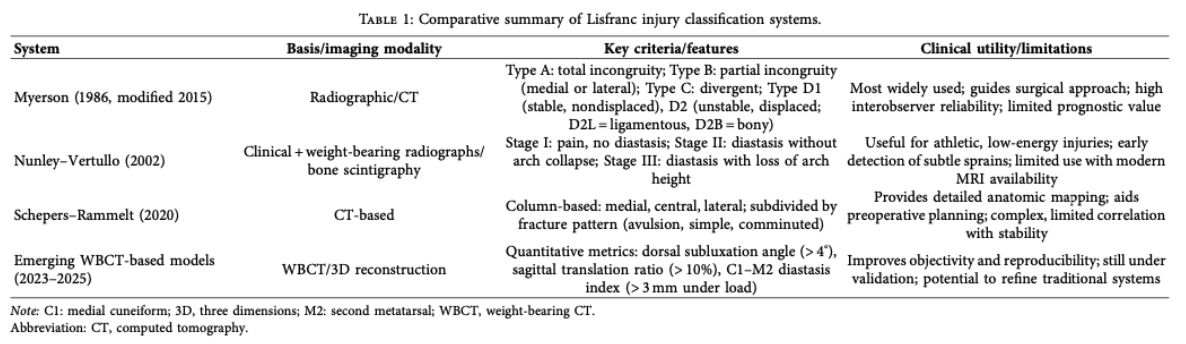
Four classification systems have been proposed for Lisfranc injuries and are detailed in Table 1. These systems help clinicians characterize injury severity and guide treatment decisions, particularly regarding surgical management. Although newer classification methods may improve the precision of clinical and surgical decision-making, further validation and development of emerging weight-bearing CT (WBCT)-based systems are needed.
Management options
Management largely depends on injury stability and displacement. Stable injuries with ≤2 mm diastasis between the second metatarsal and medial cuneiform may be managed conservatively, typically with relative rest and non-weight-bearing immobilization for 4–6 weeks, followed by gradual reloading.
For >2 mm displacement at the C1–M2 articulation, surgical fixation is generally considered. Similarly, ≥15° of sagittal talometatarsal angulation may indicate instability and support surgical management. Surgery may also be considered when symptoms persist, or joint malalignment remains.
However, these thresholds are largely clinically driven, and further research is needed to establish standardized criteria for treatment selection.
Surgical management
Common surgical approaches include open reduction and internal fixation (ORIF), arthrodesis, bridge plating, flexible fixation, and percutaneous techniques. Regardless of the technique used, the primary goal is to restore and maintain anatomic alignment, which is strongly associated with better clinical outcomes.
Rehabilitation
Following Lisfranc surgery, patients typically remain non-weight-bearing for 6–8 weeks, followed by a gradual progression to weight-bearing. Transition to normal footwear is generally expected around 12 weeks, when physiotherapy can focus on gait retraining, edema management, and restoring joint mobility.
A phase-based rehabilitation protocol has been proposed and is detailed in the table below.
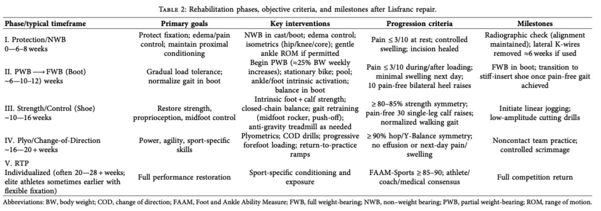
Notably, normalization of strength, balance, and hop symmetry threshold are prerequisites for phase IV, where more athlete-focused programs can restart. Interestingly, the rehabilitation program should focus on restoring normal forefoot rocker and push-off mechanics that are commonly impaired after Lisfranc injury. Rocker mechanism refers to the rolling over the metatarsophalangeal joint to allow force transmission during late stance.
Returns to ambulation and sport
Anatomic alignment is a key predictor of recovery. Weight-bearing is typically resumed after 6–8 weeks of immobilization, with most patients returning to normal footwear around 3 months. Full recovery may take up to 6 months, depending on injury severity.
Rehabilitation should address the impaired foot rocker mechanism, with progressive loading targeting foot intrinsic muscles, tibialis posterior, plantarflexor strength, and forefoot function.
Before return to sport, consider:
Pain-free sport-specific drills
- <10% side-to-side edema difference
- ≥90% symmetry on Y-Balance and single-leg hop tests
- ≥90% plantarflexion strength symmetry
- FAAM-Sports ≥85–90
Across systematic reviews and athletic case series, return to sport is typically reported around 16–28 weeks, although timelines vary with injury severity and treatment.
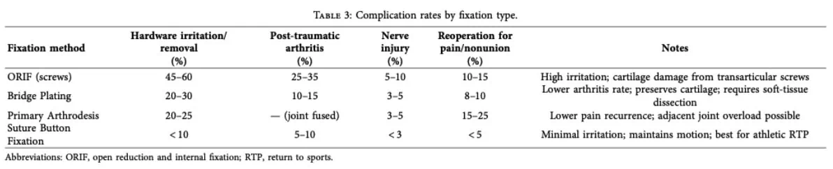
Non-operative treatment and ORIF can provide comparable outcomes when the injury is stable and anatomic alignment is maintained. Compared with arthrodesis and suture-button fixation, ORIF may be associated with poorer outcomes, potentially due to higher rates of hardware irritation and cartilage damage.
Suture-button fixation has shown promising RTS outcomes, with a mean return to sport of 17 weeks and 100% RTP reported in a small cohort of athletes. However, these findings should be interpreted cautiously given the limited evidence.
Residual performance deficits are common after Lisfranc injuries, particularly in athletes with ≥2 mm of residual displacement, reinforcing the importance of achieving and maintaining anatomic alignment.
Complications of delayed, missed, or inadequate treatment
Missed Lisfranc injuries and inadequate fixation, particularly when residual displacement is ≥2 mm, have been associated with poorer functional outcomes and lower rates of return to play. Persistent malalignment may contribute to post-traumatic osteoarthritis and, in some cases, require surgical fixation or arthrodesis of the first three tarsometatarsal joints.
Delayed or inadequate treatment may also result in persistent foot deformity, including planovalgus or cavus alignment and forefoot abduction or adduction. When surgical treatment is delayed beyond approximately 6 weeks, management can become more challenging due to ligamentous scarring and adaptive changes in the surrounding soft tissues and musculature.
Potential complications following surgical treatment are summarised in Table 3.
Questions and thoughts
The high rate of missed Lisfranc injuries should raise awareness among healthcare professionals, particularly given the potential consequences of delayed diagnosis. With approximately 30% of diagnoses of Lisfranc injuries initially missed, this represents an important gap in clinical practice. For physiotherapists, this also highlights the importance of strengthening our diagnostic skills as direct access to physiotherapy continues to expand worldwide. Developing strong differential diagnostic reasoning is therefore essential to identifying conditions that require further medical investigation or management.
A high-energy rotational injury involving a plantarflexed foot should immediately trigger a broad differential diagnosis. The initial clinical assessment should focus on identifying serious conditions and those requiring medical management. In the foot and ankle, fracture is one of the primary diagnoses to exclude. Physiotherapists should consider fractures of the navicular, cuneiforms, cuboid, and talar dome, particularly when the mechanism and clinical presentation are suggestive. Vascular compromise should also be considered, particularly following high-energy trauma, with conditions such as acute compartment syndrome or arterial occlusion requiring urgent medical or surgical assessment.
The Ottawa Ankle Rules can support fracture screening, given their high sensitivity. However, a negative screening result should not end the clinical reasoning process when a Lisfranc injury is suspected. These injuries can be missed on conventional radiographs, making a thorough midfoot examination essential. Findings such as plantar ecchymosis, asymmetry of the medial arch, deformity around the first tarsometatarsal joint, localised swelling, a positive piano-key test, and pain with first metatarsal mobilisation should increase clinical suspicion and support referral for appropriate weight-bearing imaging.
Talk nerdy to me
Narrative reviews are subject to several potential sources of bias, mainly because the search, selection, and interpretation of evidence are often less standardized than in systematic reviews.
First, selection bias is an important consideration. In this case, the review was conducted by three researchers from the same clinical institution, which may increase the likelihood of shared clinical perspectives or assumptions. Their pre-existing beliefs could also contribute to confirmation bias, whereby evidence supporting their views is preferentially identified, selected, or emphasized.
The authors report conducting “random searches” on Google Scholar, but limited information is provided regarding the exact search strategy or search terms. This makes it difficult to assess how comprehensive the search was and raises the possibility of search and citation bias. Similarly, positive findings may be preferentially included or emphasized, although this should be distinguished from publication bias, which occurs when studies with certain results are more likely to be published in the first place.
Another limitation is interpretation bias. The reader is not directly presented with the raw data from the original studies, but rather with the authors’ interpretation of those findings. Furthermore, Physiotutors reviews such as this one introduce an additional layer of interpretation that may influence the overall conclusions. So, how can these limitations be addressed? PRISMA can improve transparency in reporting, but it was primarily designed for systematic reviews and does not specifically assess the quality or risk of bias of narrative reviews. SANRA, on the other hand, was specifically developed to assess the methodological and reporting quality of narrative reviews, although it is not a conventional risk-of-bias tool.
Finally, when a narrative review aims to establish expert consensus, a Delphi consensus process can provide a more structured approach. By collecting and refining opinions across multiple rounds and allowing experts to reconsider their responses, Delphi methodology can reduce the influence of individual opinions and promote a more transparent consensus. However, it cannot completely eliminate bias, particularly if the expert panel itself is not representative.
Take-home messages
- Think Lisfranc: Around 30% of Lisfranc injury diagnoses are initially missed. Midfoot pain following a twisting injury to a plantarflexed foot should raise suspicion rather than being labelled a simple ankle sprain.
- Screen for serious pathology first: Consider fractures of the navicular, cuneiforms, cuboid, or talar dome, as well as vascular compromise. The Ottawa Ankle Rules can help screen for fractures but should not replace a thorough clinical assessment.
- Look for key clinical signs: Plantar ecchymosis, a positive piano-key test, pain with first tarsometatarsal joint stress, and asymmetry of the medial arch can increase suspicion of a Lisfranc injury.
- Don’t rely on standard X-rays alone: Weight-bearing radiographs are preferred when a Lisfranc injury diagnosis is suspected. CT can help assess bony displacement, while MRI may be useful for evaluating ligamentous injury.
- Management depends on stability: Stable, minimally displaced injuries may be managed conservatively, whereas unstable or significantly displaced injuries often require surgical fixation. Anatomic reduction is consistently associated with better outcomes.
- Rehabilitation is progressive: After immobilization or surgery, gradually restore weight-bearing, foot and ankle strength, and functional loading. Later-stage rehabilitation should address plantarflexor capacity and forefoot rocker mechanics.
- Return to sport should be criteria-based: Consider pain, swelling, strength, balance, and sport-specific function rather than relying on time alone. Recovery may take several months.
- Don’t underestimate Lisfranc injuries: Delayed diagnosis can contribute to persistent symptoms, deformity, post-traumatic osteoarthritis, and more complex treatment.
- Keep the evidence in context: These recommendations are based on a narrative review and should be interpreted as clinical guidance rather than guideline-level evidence.
Reference
Additional references
Complete Anatomy, 3D4Medical. (2026). [Computer software]. Elsevier.
21 OF THE MOST USEFUL ORTHOPAEDIC TESTS IN CLINICAL PRACTICE
We have compiled a 100% Free E-Book containing 21 of the hands-down most useful orthopedic tests per body region, guaranteed to help you reach a proper diagnosis today!