Ankle/Foot Assessment

Learn
Introduction & Epidemiology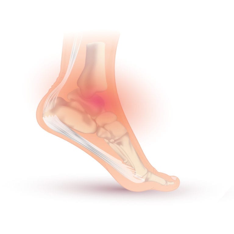
First and foremost let’s talk a little bit about anatomy. The ankle joint consists of two different joints:
- The talocrural joint, which is a hinge joint allowing the two primary movements in the sagittal plane: plantar flexion and dorsiflexion
- The subtalar joint is considered a hinge joint as well but its rotational axis runs obliquely. The possible movements at this joint are: inversion (a combination of plantar flexion, adduction and supination) and eversion (a combination of dorsiflexion, abduction and pronation).
Note: In the US nomenclature inversion is referred to as supination and eversion as pronation.
The one-year prevalence for ankle- and foot injuries seen in general practice is estimated at 9.2% and 9.4% respectively (Picavet et al. 2003).
These can be traumatic and atraumatic in nature.
Common conditions for both categories are:
Traumatic:
- Ankle sprain with or without syndesmosis lesion
- Ankle fracture
- Achilles tendon rupture
Atraumatic:
- Tendinopathies/ Plantar fasciitis
- Hallux valgus/rigidus
- Osteoarthritis of the ankle
Course
Not much can be said about the general course of ankle and foot pain and detailed prognostics are discussed in the individual chapters. Furthermore, it’s unclear which specific prognostic factors influence the course of ankle and foot injuries.
Prognostic factors (Artus et al. 2017)
The following factors are related, but not limited to, the course of ankle and foot pain:
- widespread pain
- high functional disability
- somatization
- high pain intensity
- presence of previous pain episodes
Where available, specific prognostic factors, as well as risk factors, are discussed in the individual units.
Red flags
There are several specific pathologies that count as red flags. These are:
Fractures
The Ottawa Ankle Rules are a great tool to assess for fractures. We discuss them more in detail in the ankle sprain unit.
Signs and symptoms include:
-
Immediate and severe pain
-
Swelling
-
Bruising
-
Localized tenderness upon palpation
-
Inability to bear weight
-
Deformity, especially in case of a dislocation
Osteochondral lesions, osteophytes
Signs and symptoms include:
- Persisting synovitis
- Stiffness
- Ventral impingement causes limitation in dorsi- and plantarflexion
Osteochondritis dissecans
More often seen in young individuals. Can be post-traumatic.
Signs and symptoms are:
- Intermittent pain
- Swelling
- Clicking in the joint
- Moderate synovitis
Sinus Tarsi Syndrome
Signs and symptoms include:
- “Giving-way” feeling
- Tenderness 2cm anteriorly and distally from the tip of the lateral malleolus
Muscle- or tendon tears
The patient is unable to contract the muscles around the ankle
WHAT TO LOOK FOR TO PREVENT HAMSTRING, CALF & QUADRICEPS INJURIES

Basic Assessment
Depending on the outcome, your basic assessment can give you the following info:
1) Limitations in range of motion and their end-feel can guide structural assessment (e.g. bone to bone=osteoarthritis, empty=tendinopathy due to pain)
It’s best to start with Active Range of Motion Assessment:
Standard values for the range of motion in different directions are as follows:
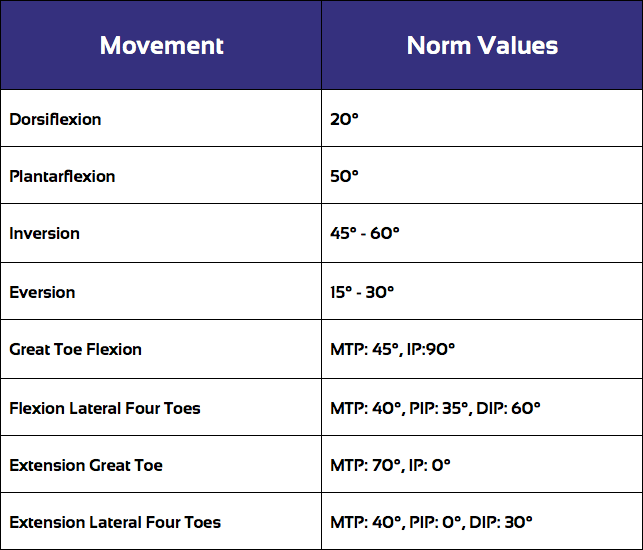
AROM assessment is then typically followed by Passive Range of Motion Assessment (PROM) which you can watch by a click on the following video:
During PROM assessment, it’s important to compare the range of motion as well as the end-feel of the affected ankle with the unaffected side.
Don’t forget that inspection and palpation can also give you clues. Think swelling, hematoma, warmth, pain or tenderness. These can guide you in identifying which structures may be affected.
Specific Pathologies in the Ankle & Foot
There are several pathologies that are commonly seen in the ankle & foot area. For more information, click on the respective pathology (content will be added in the near future):
- Syndesmosis Injury
- Ankle Impingement
- Tarsal Tunnel Syndrome
- Inversion Trauma and Lateral Ankle Sprain
- Achilles Tendon Rupture
- Achilles Tendinopathy
- Plantar Fasciitis
References
Accredited online physiotherapy courses
- Built by the experts at Physiotutors
- Best price per CEUs/CPD Points
- Accredited in the Netherlands, Belgium, Germany, USA, UK, & Australia
- Learn anywhere, any time, and at your own pace!


Download the free Physiotutors app now!


