Lateral Epicondylalgia

Body Chart
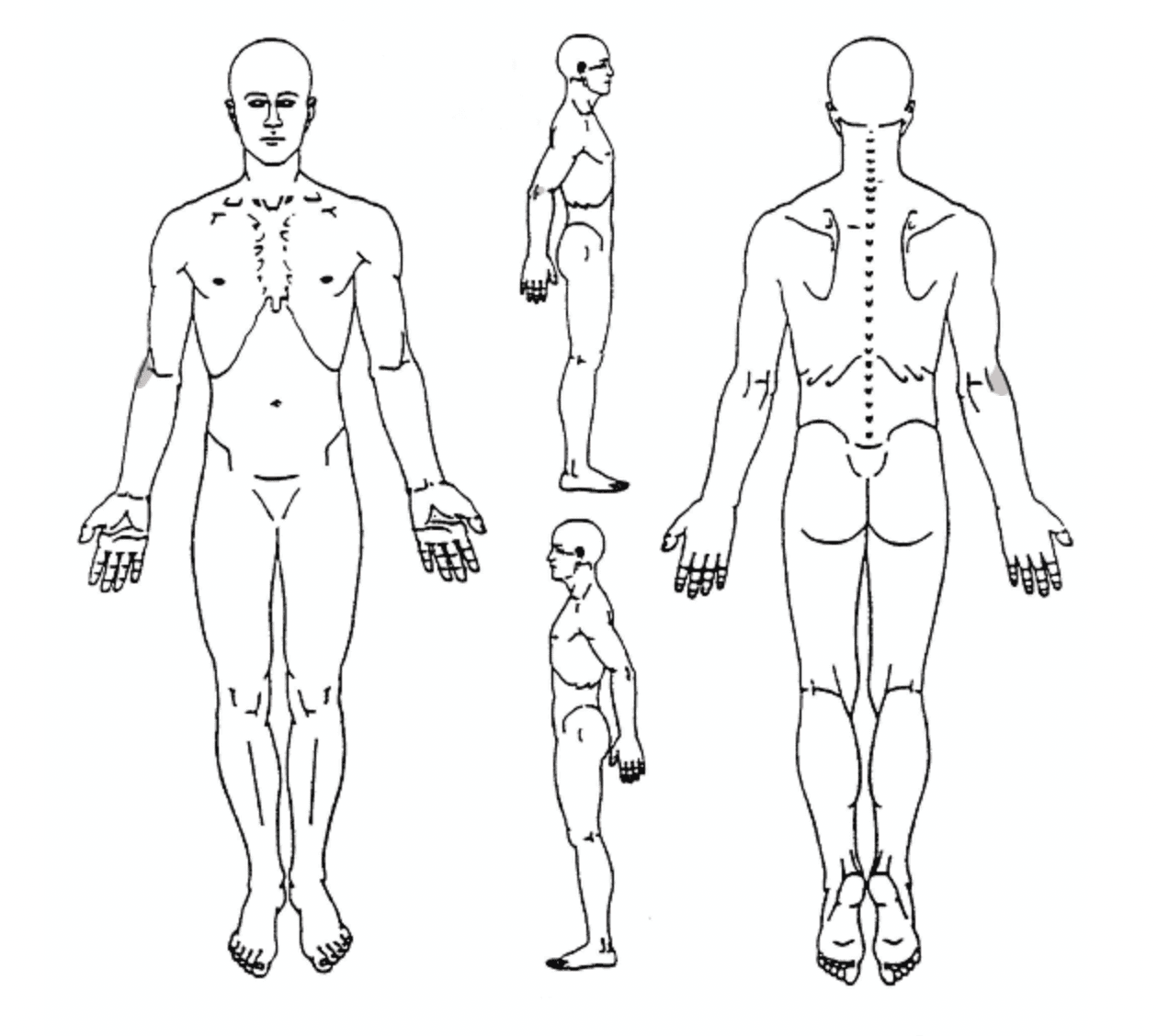
- Localized around the lateral elbow/humeral epicondyle
Background Information
Patient Profile
- Between 20-50 years old
- Female = male
- Dominant side affected
Pathophysiology
Result from overuse of the extensor carpi radialis brevis (ECRB) muscle by repetitive microtrauma resulting in primary tendinosis of the ECRB, with or without involvement of the extensor digitorum communis. Fibroplastic tissue and vascular invasion define a degenerative process characterized by an abundance of fibroblasts, vascular hyperplasia, and unstructured collagen. Pain in Lateral Epicondylalgia is explained by ingrowing free nerve endings and blood vessels into the degenerated tendon.
Course
In the descriptive epidemiology study by Sanders et al. (2015) 50% of patients had only 1-2 visits for lateral elbow tendinosis and 74% were no longer seeking care after three months of their initial diagnosis. However, of the 18% of patients who still received care after six months, the median duration of care was 844 days. Eventually 12,3% of these patients went on to have surgery with a mean time to operation of about nine months after symptom onset.In a prognostic cohort study of Smidt et al. (2006) 89% of patients reported improvement in pain at one year follow-up.
History & Physical Examination
History
Long history, patients tend to ignore early symptoms; Visit to the doctor rather late; Patients describe trauma or repetitive unilateral tasks at work, during ADLs or sport; pain of gradual onset
- Pain around lateral epicondyle
- Radiating up and down
- Stiff
- Stinging
- Loss of strength
Physical Examination
Inspection & Palpation
Palpation of lateral humeral epicondyle on pain provocation
Functional Assessment
Patients can demonstrate pain aggravating movements (carrying, gripping)
Special Testing
Other physical examination is obsolete
Differential Diagnosis
- Radiculopathy
- LCL injury
- Biceps/triceps tendinopathy
- Radius fracture
- Cervical Radicular Syndrome
Treatment
Strategy
Avoid provoking activities. Education. Progressively increase strength of wrist extensors
Interventions
- Rest: avoid painful activities, pain dictates load
- Educate on: condition, workplace ergonomics, self-management
- No clear evidence whether concentric or eccentric training is superior.
- MT: MWM appear effective, Mill’s manipulation
Literatuur
- Bisset, Leanne M., and Bill Vicenzino. “Physiotherapy management of lateral epicondylalgia.” Journal of physiotherapy 61.4 (2015): 174-181.
- Bot SD, Van der Waal JM, Terwee CB, Van der Windt DA, Schellevis FG, Bouter LM et al. Incidence and prevalence of complaints of the neck and upper extremity in general practice. Ann Rheum Dis 2005a;64:118-23
- Macfarlane, G.J., I.M. Hunt, and A.J. Silman, Role of mechanical andpsychosocial factors in the onset of forearm pain: prospective population based study. BMJ, 2000. 321(7262): p. 676-9.
- Nagrale, Amit V., et al. “Cyriax physiotherapy versus phonophoresis with supervised exercise in subjects with lateral epicondylalgia: a randomized clinical trial.” Journal of Manual & Manipulative Therapy 17.3 (2009): 171-178.
- NHG Standaard Epicondylitis Lateralis, NHG, 2009
- Sorgatz, H., Repetitive strain injuries. Forearm pain caused by tissue responses to repetitive strain. Orthopade, 2002. 31(10): p. 1006-14.
- Vaquero-Picado A1, Barco R1, Antuña SA1. Lateral epicondylitis of the elbow. EFORT Open Rev. 2017 Mar 13;1(11):391-397
- Verhagen AP, Alessi J. Evidence based diagnostiek van het bewegingsapparaat. 2014Walz, J. S. Newman, G. P. Konin, and G. Ross, Epicondylitis: Pathogenesis, Imaging, and Treatment, RadioGraphics, January 1, 2010; 30(1): 167 – 184. Level of Evidence: 2C