Lateral Ankle Sprain | Diagnosis & Treatment
Clinical Presentation & Examination
Diagnosis
Risk Factors
Sports with high-speed collisions, artificial surfaces, unlevel ground, and high-torque cutting and jumping forces, which can cause dorsiflexion and external rotation of the foot in relation to the ankle and tibia, are more likely to cause syndesmotic injuries (eg, football, soccer, basketball, rugby, skiing, hockey) (Hunt et al., 2013).
Clinical Evaluation
The diagnosis of syndesmotic injuries includes a thorough history and physical examination, as well as imaging tests such as X-rays, CT scans, MRI scans, or ultrasound to confirm the presence of a syndesmotic injury and to rule out other potential causes of pain and instability in the ankle joint.
Physical Examination
During the physical examination, the physiotherapist will assess the patient’s range of motion, stability, and pain in the affected joint. Special tests, such as the squeeze test or the external rotation stress test, may be performed to further evaluate the integrity of the syndesmotic complex. When a syndesmotic injury is suspected, imaging is recommended (van Dijk et al., 2015).
Tenderness on palpation of the syndesmosis ligaments is the most sensitive test while the squeeze test is the most specific (Sman et al., 2015). Both being positive results in a high probability of injury to the syndesmosis ligaments.
In terms of classification, many models have been proposed. Currently, there is no consensus on what classification to use. As a broad guide, you can divide them into stable and unstable joints, and isolated and non-isolated injuries.
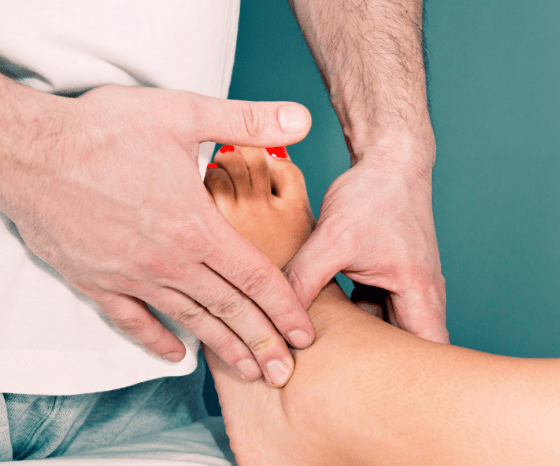
Palpation syndesmosis ligaments:
Squeeze test:
Other tests you might consider are:
The Cotton test:
The fibular translation test:
Heel thump test:
Imaging Tests
In terms of imaging, X-rays can provide information about the position and alignment of the ankle joint, while CT scans and MRI scans can provide more detailed information about the ligaments and surrounding soft tissues. Magnetic resonance imaging has excellent sensitivity and specificity for visualizing syndesmotic injuries, although an arthroscopy remains the gold standard. Diastasis can be present, resulting in an unstable joint (van Dijk et al., 2015). To assess this appropriately, a unilateral weight-bearing film is preferred. However, patients may not tolerate this in the early phase (Lin et al., 2006).
Be aware of a potential Maisonneuve fracture. This is a commonly missed fracture of the proximal fibula that might occur during ankle trauma (Taweel et al. 2013)
LEVEL UP YOUR ROTATOR CUFF DISORDER KNOWLEDGE – FOR FREE!