

Research
Ankle/Foot
August 18, 2026
Foot and Ankle Biomechanics in Patellofemoral Pain: What Does the Evidence Tell Us?
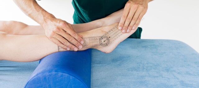
This systematic review with meta-analysis investigated foot and ankle biomechanical characteristics associated with patellofemoral pain and found strong evidence for greater navicular drop in individuals with patellofemoral pain
Other biomechanical findings were inconsistent, with greater rearfoot eversion observed only during some functional tasks and no consistent differences during walking, running, or single-leg squat
Clinical tests assess different aspects of foot mechanics and should not be interpreted interchangeably. Their relevance should ultimately be determined by whether the identified biomechanical feature relates to the individual patient’s symptoms and clinical presentation
Introduction
Patellofemoral pain is a common musculoskeletal condition affecting young and physically active individuals. It is characterized by pain around or behind the patella that is typically aggravated by activities such as jumping, squatting, and stair climbing. One approach to understanding patellofemoral pain involves a biomechanical paradigm. Systematic reviews and meta-analyses have reported an association between patellofemoral pain and dynamic knee valgus, which refers to the medial displacement of the knee during weight-bearing movements. This altered knee position may result from suboptimal alignment or movement patterns within the lower-limb kinetic chain. In particular, excessive hip adduction and internal rotation, as well as rearfoot eversion, have been identified as movements associated with dynamic knee valgus.
Previous research has demonstrated an association between increased rearfoot eversion and patellofemoral pain in female runners. However, no systematic review has comprehensively investigated the range of biomechanical factors that may contribute to patellofemoral pain. Therefore, this systematic review and meta-analysis aimed to evaluate the contribution of ankle biomechanics in patellofemoral pain. In addition, the study investigated potential sources of heterogeneity across studies to improve the interpretation of the existing evidence and inform clinical reasoning and future research.
Methods
This study is a systematic review with meta-analysis.
Inclusion criteria
Case-control studies, prospective studies, and clinical trials investigating ankle biomechanics between participants with and without patellofemoral pain. Studies were eligible for inclusion if they met the following criteria:
- Human participants
- Participants diagnosed exclusively with patellofemoral pain
- Assessment of lower-extremity biomechanics, including muscle strength, kinematics, and range of motion
- Functional assessment of the ankle and foot, including measures such as the navicular drop test and foot mobility
Exclusion criteria
Review articles, conference proceedings, letters, and symposium abstracts were excluded. Studies involving participants with other musculoskeletal conditions, studies that did not assess distal joint biomechanics, and studies without a control group were also excluded.
Extracted data
Data on publication characteristics and participant demographics, including age, sex, body weight, height, and physical activity profile, were extracted. Baseline clinical characteristics were also recorded, including symptom duration, pain intensity, unilateral or bilateral symptoms, and physical function.
Means and standard deviations were extracted for both the control and patellofemoral pain groups. For prospective studies and clinical trials, baseline data from the pain-free and patellofemoral pain groups were extracted; post-intervention or post-assessment data were not included in the analysis.
Methodological quality assessment
The methodological quality of the included studies was assessed using standardized, study-design-specific tools. The Downs and Black checklist was used to evaluate methodological quality on a 16-point scale. Studies scoring <9 points were considered to be of low quality, those scoring 10–11 points were considered to be of moderate quality, and those scoring ≥12 points were considered to be of high quality.
Data analysis
Studies were combined in a meta-analysis when at least two studies reported the same outcome. Muscle strength, range of motion (ROM), and kinematic outcomes were analyzed separately according to the movement and task assessed. When data were reported for different subgroups or phases of a movement, they were combined using weighted means and standard deviations based on sample size.
A random-effects model was used. Effects were reported as standardized mean differences (SMD) using Hedges’ g, with 95% confidence intervals. Effect sizes were classified as small (≤0.59), medium (0.60–1.19), or large (≥1.20). Statistical significance was set at p < 0.05. Heterogeneity was assessed using I², with values >25% and >50% indicating moderate and high heterogeneity, respectively.
Subgroup analyses were performed when sufficient data were available, based on factors such as sex or assessment method. Leave-one-out sensitivity analyses were also performed to determine whether individual studies influenced the results.
The overall level of evidence was classified as strong, moderate, limited, very limited, or conflicting, based on the number and quality of studies, statistical significance, and heterogeneity of the results.
Results
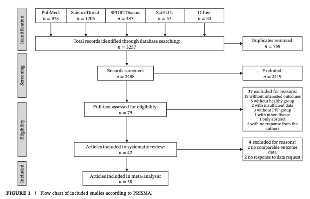
A total of 42 studies were included in the review. Due to incomplete data or methodological limitations, four studies were excluded from the meta-analysis. Of the included studies, 41 were case-control studies, and one was prospective. Overall, 2,314 participants were included, with a predominance of females (70%). Participants were aged 18–35 years, with BMI values ranging from 19 to 27 kg/m². Symptom duration, pain intensity, and functional status varied considerably across participants and were considered important potential sources of heterogeneity.
Methodological assessment
Of the included studies, 35.6% were classified as high quality, 57.1% as moderate quality, and 7.1% as low quality. Notably, none of the studies met the external validity criterion of the Downs and Black checklist. In addition, no studies reported assessor blinding or accounted for potential confounding factors.
Rearfoot eversion
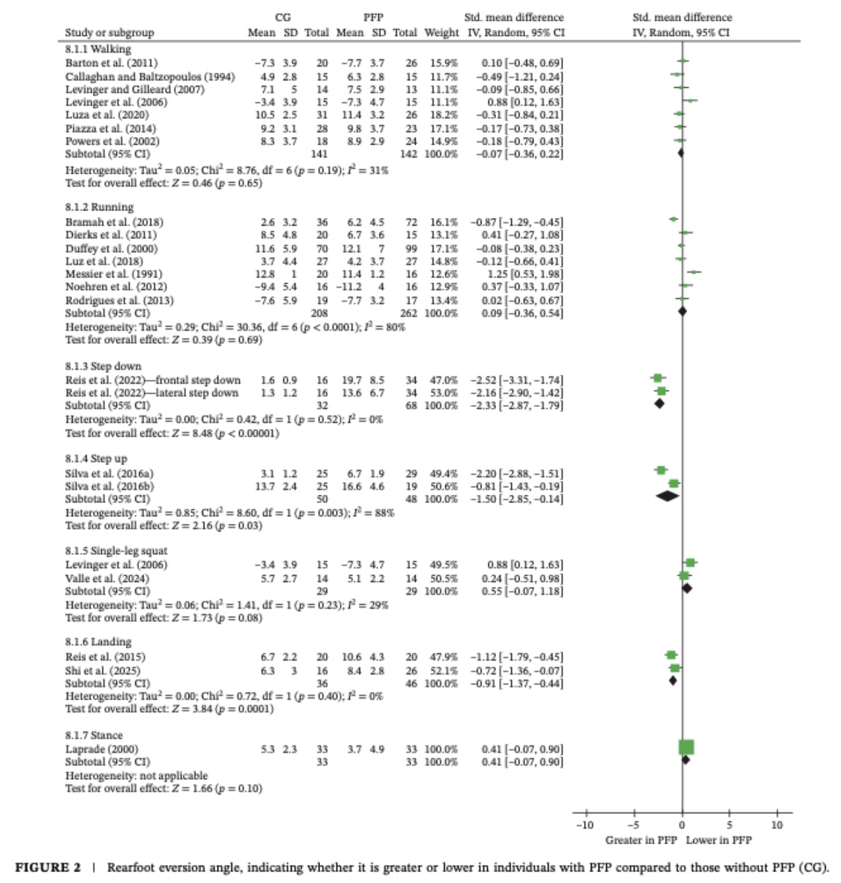
No significant between-group differences in ankle biomechanics were observed in patellofemoral pain during walking, running, or single-leg squatting. However, the patellofemoral pain group demonstrated greater rearfoot eversion during landing, step-up, and step-down tasks. These findings should be interpreted with caution, as only two studies contributed to the landing analysis, while the step-up and step-down findings were based on a single study sample.
After the leave-one-out analysis, in which each study was removed one at a time to assess its influence on the results, the findings remained non-significant for both walking and running. Overall, the evidence was considered conflicting for walking, running, and single-leg squat, and very limited for landing, step-up, and step-down tasks.
Ankle Dorsiflexion ROM assessed by kinematics
No significant between-group differences were observed during walking, running, or landing. The evidence for these outcomes was considered conflicting. Greater ankle dorsiflexion was observed in the patellofemoral pain group during the step-down task. However, this finding was based on only two studies, with a single-participant sample, and should therefore be interpreted with caution.
Subgroup analyses exploring potential sources of heterogeneity did not change the results. Similarly, although the leave-one-out analysis reduced heterogeneity to 0%, the overall findings remained unchanged.
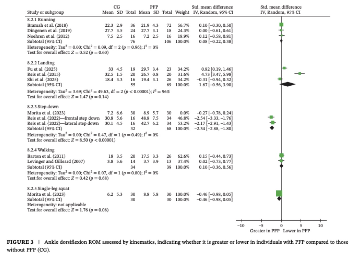
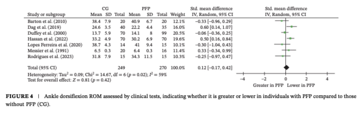
Ankle Dorsiflexion ROM using clinical tests
No significant between-group differences were found in ankle dorsiflexion range of motion (ROM) during clinical tests. Analyses exploring the impact of study heterogeneity did not alter these findings.
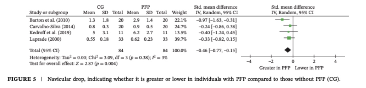
Navicular drop
Based on pooled data from four studies including 168 participants, the patellofemoral pain group demonstrated greater navicular drop than the control group. Heterogeneity was low (I² = 3%). Therefore, this finding was considered to have a strong level of evidence.
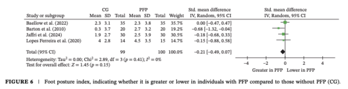
Foot posture index
No significant between-group differences were found in foot pressure index. Heterogeneity was negligible, suggesting consistent findings across the included studies.
Plantar pressure
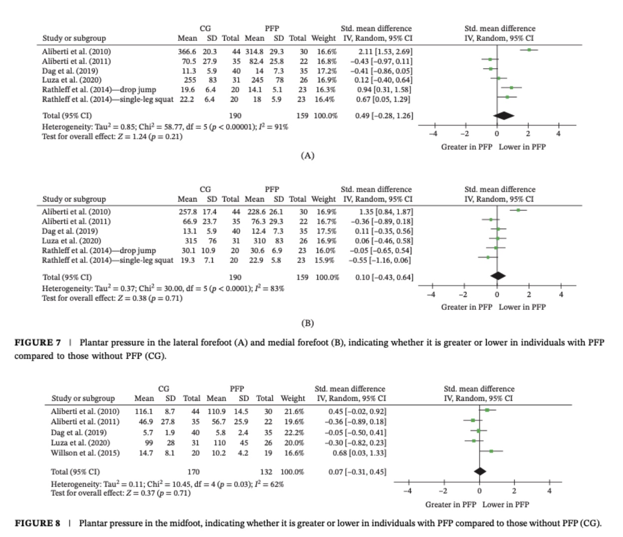
No significant between-group differences were observed across the three foot compartments (hindfoot, midfoot, and forefoot) for either medial or lateral pressure distribution. Substantial heterogeneity was observed, suggesting considerable methodological and/or clinical variability across the included studies. Therefore, the level of evidence was classified as conflicting.
Muscle strength
The authors were unable to perform a meta-analysis of muscle strength due to substantial differences in the muscle groups assessed and testing methods, which prevented meaningful pooling of the data.
Questions and thoughts
Greater navicular drop was consistently observed in individuals with patellofemoral pain. Greater rearfoot eversion was also reported during landing and step-down tasks. These findings suggest that altered distal foot mechanics may contribute to patellofemoral pain and provide some support for the biomechanical paradigm of patellofemoral pain. However, findings across other biomechanical measures were inconsistent.
It could be expected that symptomatic individuals would demonstrate broader alterations in ankle biomechanics in patellofemoral pain. If this were the case, differences in foot pressure distribution might also be expected between patellofemoral pain and asymptomatic participants. However, no significant differences were observed in foot pressure outcomes.
The findings regarding rearfoot eversion and ankle dorsiflexion also raise questions. Greater rearfoot eversion was observed during landing and step-down tasks only; no differences were found for walking, running, or single-leg squat. Greater ankle dorsiflexion was reported during step-down. This may appear counterintuitive, as rearfoot eversion should be observed more consistently during functional tasks; furthermore, reduced ankle dorsiflexion has been associated with greater dynamic knee valgus during the step-down task. Therefore, one might expect individuals with patellofemoral pain to demonstrate reduced rather than increased ankle dorsiflexion.
Interestingly, several of the included measures appear to assess related aspects of foot pronation. Rearfoot eversion, ankle dorsiflexion, foot pressure distribution, and navicular drop may all reflect different components of distal lower-limb mechanics. However, these measures are not interchangeable and may be influenced by the assessment method, task demands, and clinical context. This could partly explain the inconsistent findings observed across studies.
Talk nerdy to me
The findings of this meta-analysis may appear contradictory at first glance. But before concluding that the biomechanical contribution of the foot and ankle to patellofemoral pain is inconsistent, we should ask a more fundamental question: are these clinical tests actually measuring the same mechanical construct?
The objective of the review was to determine whether biomechanical and clinical parameters of the foot and ankle differ between individuals with and without patellofemoral pain. This question is highly relevant clinically, because we often use these tests to confirm or refute a biomechanical hypothesis. However, the tests included in this review assess different aspects of foot and ankle mechanics, often under very different conditions.
Let’s take a closer look at the different tests.
Navicular drop test: The navicular drop test assesses the change in navicular height between a standardized position and relaxed standing. It therefore provides an estimate of the amount of medial longitudinal arch deformation under load and can be considered an indirect measure of foot pronation mobility. The test is simple and clinically useful, but it is performed in a static, standardized position. It therefore provides limited information about how the foot behaves dynamically. A greater navicular drop may indicate greater pronation structural mobility, but it does not tell us whether this movement is actually problematic or contributes to the patient’s symptoms.
Foot Posture Index (FPI): The FPI assesses the overall position of the foot in standing, providing an indication of whether the foot has a more pronated or supinated posture. However, it remains a static assessment and cannot determine why this posture is present or whether it influences movement during functional activities. It is also subject to examiner-dependent measurement and positioning. In addition, because the assessment is performed in a standardized standing position, the patient’s posture may be influenced by the instructions or positioning provided by the examiner. This introduces the possibility of measurement or examiner-related bias, although it should not necessarily be assumed that patients consciously alter their posture to meet examiner expectations.
Plantar pressure: Plantar pressure provides information about how mechanical load is distributed across the foot during a specific task. However, plantar pressure is not a direct measure of foot pronation. It can be influenced by multiple factors, including pain, compensations, walking or running strategy, footwear, speed, task demands, and individual movement patterns. Importantly, the studies included in the review assessed plantar pressure during very different activities, such as walking, running, jumping, or stair-related tasks. These differences in testing conditions could explain part of the substantial heterogeneity observed between studies. More importantly, differences in plantar pressure do not necessarily indicate differences in foot posture or pronation.
Rearfoot eversion: Rearfoot eversion provides a more direct measure of rearfoot motion and is commonly used as a proxy for rearfoot pronation. However, its interpretation is highly dependent on the task being performed. Rearfoot eversion during quiet standing is not necessarily equivalent to rearfoot eversion during running, landing, or a step-down. The magnitude, timing, and rate of the movement may all be clinically relevant. Therefore, simply finding greater eversion does not necessarily mean that the movement is pathological or causally related to patellofemoral pain.
This raises an important clinical point: a difference between groups does not automatically mean that the measured variable is clinically meaningful. A test can demonstrate excellent reliability and still have limited clinical relevance if it does not capture the movement or loading strategy that is relevant to the patient’s symptoms.
Another important consideration is the distinction between impairment and contribution. Finding greater navicular drop or rearfoot eversion in a patient with patellofemoral pain does not demonstrate that these factors caused the pain. They may be associated features, compensatory strategies, or simply characteristics that coexist with patellofemoral pain. The clinical value of a test therefore depends not only on whether it distinguishes patients from asymptomatic individuals, but also on whether the results help us make clinically meaningful decisions.
Finally, these tests may be better viewed as different pieces of the same puzzle rather than interchangeable measures of foot pronation. Navicular drop assesses arch deformation, the FPI evaluates static foot posture, plantar pressure reflects load distribution, and rearfoot eversion captures rearfoot motion. Although these measures are related, they represent distinct aspects of foot mechanics. Ultimately, the key clinical question is not simply whether a biomechanical alteration is present, but whether the observed alteration is relevant to the patient’s symptoms and clinical presentation.
Take-home messages
- Navicular drop was consistently greater in individuals with patellofemoral pain, with strong evidence and low heterogeneity, suggesting an association between patellofemoral pain and increased medial arch mobility under load.
- Evidence for other foot and ankle biomechanics in patellofemoral pain patients was inconsistent. Rearfoot eversion differed only during some functional tasks, while no consistent differences were found during walking, running, or single-leg squat.
- Clinical measures do not necessarily reflect dynamic foot mechanics. Navicular drop, FPI, plantar pressure, and rearfoot eversion assess different aspects of foot function and should not be considered interchangeable measures of “foot pronation.”
- A biomechanical difference does not necessarily indicate a clinically relevant impairment. The presence of greater navicular drop or rearfoot eversion does not establish that these factors cause or contribute to an individual’s patellofemoral pain.
- Context matters when interpreting biomechanical tests. Task selection, assessment method, pain, and individual movement strategies may influence findings and contribute to the heterogeneity observed across studies.
- Clinical tests should be interpreted in relation to the patient’s presentation. Rather than asking whether a patient has “abnormal” foot biomechanics, clinicians should ask whether the identified biomechanical feature is relevant to the patient’s symptoms and can inform clinical decision-making.
Reference
THE ROLE OF THE VMO & QUADS IN PFP
Watch this FREE 2-PART VIDEO LECTURE by knee pain expert Claire Robertson who dissects the literature on the topic and how it impacts clinical practice.