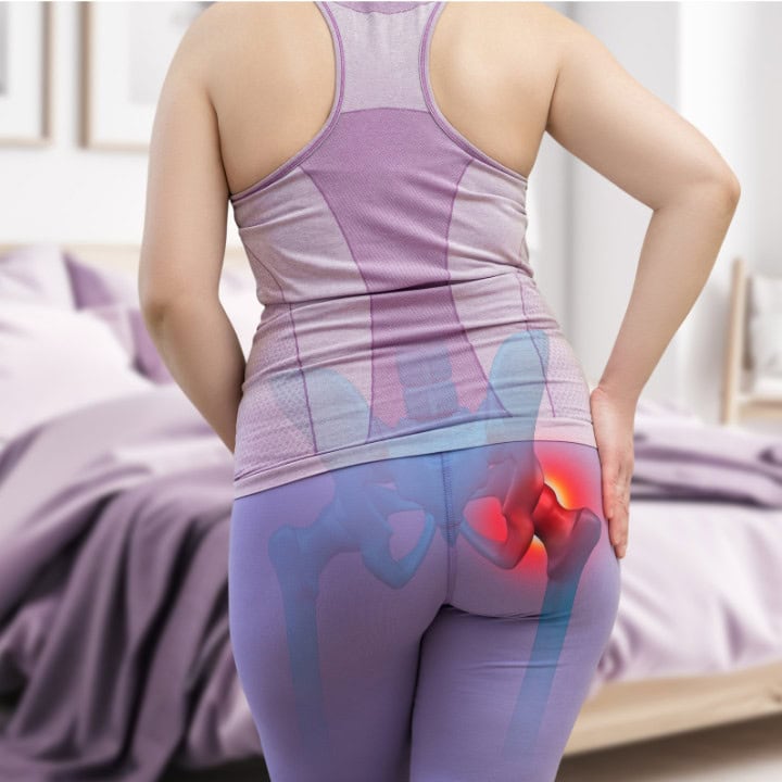
For physiotherapists working in primary care and musculoskeletal settings, the use of diagnostic imaging remains a complex and often controversial issue. While clinical guidelines offer a relatively unified stance, the realities of day-to-day practice introduce pressures, uncertainties, and system-level tensions that influence imaging decisions.
This article explores the core principles of appropriate imaging use, the divergence from best-practice recommendations, and the clinical reasoning required to navigate these decisions effectively.
The content of this post is largely based on Andrew Cuff’s work.
Clinical Guidelines: When Should Imaging Be Used?
High-quality clinical guidelines, based on systematic evidence reviews, are unequivocal in their recommendations for non-traumatic MSK conditions. Imaging should be used only when:
- There is suspicion of serious or specific pathology.
- A patient has not responded to an adequate trial of conservative management.
- The result is expected to alter clinical management.
These criteria aim to prevent unnecessary investigations and downstream effects that do not improve outcomes. Despite their clarity of these recommendations, implementation remains highly inconsistent.
Rising Imaging Rates: A Global Trend
Across healthcare systems, diagnostic imaging for MSK pain continues to rise. In the UK, for example, imaging rates increased approximately 20% between 2013 and the onset of the COVID-19 pandemic. Similar patterns are observed internationally, particularly in Australia and the United States.
A key driver post-pandemic has been the focus on a healthcare system “recovery” agenda. Targets to exceed pre-COVID imaging volumes (up to 120%) have been introduced, based on the assumption that increased rates of imaging equates to a better healthcare service. This idea unintentionally reinforces volume rather than appropriateness as a performance metric and creates a tension between policy and evidence-based care.
The fundamental question remains: Does increased imaging improve outcomes? Currently, the evidence indicates it does not. Despite higher imaging utilisation, pain-related disability, chronicity, and complexity remain unchanged.
The Key Question: Will Imaging Change What Happens Next?
One of the most clinically relevant—but frequently overlooked—questions is:
Will this imaging result change what I do?
Will this imaging result change what I do?
In many cases, imaging is performed when patients fail to improve, but without a clear plan for what would follow based on the findings. Imaging in such scenarios serves more to manage clinician uncertainty or patient expectations than to support evidence-based decision-making.
Importantly, imaging should only be pursued if a meaningful therapeutic decision—often involving an invasive intervention such as injection or surgery—is both under consideration and acceptable to the patient. If not, the rationale for imaging becomes weak.

Why Is There a Gap Between Guidelines and Practice?
Several interlocking factors contribute to the persistent gap between what guidelines recommend and what clinicians do:
- Time pressure and clinical fatigue: Short consultation windows and long patient lists reduce the bandwidth available for nuanced clinical reasoning or shared decision-making.
- Patient expectations: When patients expect imaging, clinicians often comply—especially after repeated, energy-consuming discussions in the same clinical day.
- Patient complexity: Patients are presenting with increasingly complex profiles—chronicity, multimorbidity, and lower levels of health literacy. These factors make diagnostic reasoning more difficult and increase the likelihood that clinicians default to imaging.
- Diagnostic uncertainty: Imaging is often used to rule out serious pathology, especially when the clinical picture is unclear. This use is not always justified by guidelines but is influenced by the clinician’s need for reassurance. Physiotherapists, in particular, often demonstrate a lower tolerance for clinical uncertainty and risk compared to general practitioners or other medical colleagues. While GPs regularly manage uncertainty without immediate recourse to investigations, physiotherapists may be quicker to seek imaging as a safeguard, both to reassure themselves they are not missing pathology and to reduce perceived medico-legal exposure. Compounding this is the fact that real-life patients are typically far more complex than patient profiles described in clinical guidelines. This mismatch makes it harder to map guideline recommendations directly onto the realities of clinical practice..
Professional comfort with clinical uncertainty is particularly limited in physiotherapy practice. Developing this tolerance is essential as physiotherapists increasingly take on first-contact and advanced roles in MSK care.
Physiotherapists, in particular, often demonstrate a lower tolerance for clinical uncertainty and risk compared to general practitioners or other medical colleagues.
Imaging as a Clinical Tool: Appropriate Use Cases
Imaging can be valuable—but only in the right context and while considering its limitations. Recommended use includes:
- Suspected red flag conditions (e.g., malignancy, infection, fracture).
- Specific diagnoses with clear medical treatments, such as axial spondyloarthritis or gout.
- Lack of progress after conservative treatment, where a next-step intervention (e.g., corticosteroid injection or surgical referral) is being actively considered and the patient is open to it.
In one large UK-based community MSK service seeing approximately half a million patients annually, imaging was used in only around 4% of cases—demonstrating that conservative care can usually proceed without it.
Sciatica and Early Imaging: A Clinical Dilemma
Severe acute sciatica presents a well-recognised grey area in physiotherapy. Early imaging in these cases may help inform time-sensitive decisions regarding epidural injection or surgical intervention. Yet early scanning also risks reinforcing a biomedical narrative and initiating premature interventions.The appropriate strategy is to frame the decision through shared reasoning. If the patient is open to invasive options, imaging may be warranted. If not, conservative management can proceed without it.
Patient Expectations: Who Really Wants the Scan?
It is often assumed that patients push hardest for imaging. However, qualitative research indicates that imaging is most commonly introduced by the clinician, not requested by the patient. Patients report that what they truly value is a clear understanding of their condition. When that is provided, many are content to proceed without imaging—highlighting the value of communication over investigation.
The way imaging is introduced by the clinician can strongly influence patient thinking. When a clinician suggests a scan, even tentatively, patients often interpret it as a sign that something more serious may be wrong. A thought that might not have existed before suddenly takes root: “If the clinician thinks a scan could help, does that mean they are worried?” This shift can turn a previously neutral stance into one of expectation or even insistence on imaging. In effect, the clinician’s suggestion reframes the patient’s perception of their own problem and plants doubt that non-imaging approaches might miss something important.
This dynamic underscores the need for clinicians to be careful in how and when imaging is mentioned. Introducing it casually or as an afterthought can create demand where none previously existed, making shared decision-making more difficult. Clear, confident explanations about the limits and appropriate role of imaging are essential to prevent this cascade.
Ortopedisk fysioterapi av ryggraden
Improve your ability to manage patients with spinal and sacroiliac joint complaints!

What Imaging Can and Cannot Do
The most important limitation of imaging is straightforward: It cannot identify pain.
While useful for excluding pathology, imaging struggles to explain most non-specific MSK symptoms. Findings often do not correlate with pain severity or functional limitation. Yet patients—and sometimes clinicians—continue to seek a definitive diagnosis through scans.
Even when imaging doesn’t change treatment, patients often feel reassured. The validation of their symptoms, and the exclusion of serious pathology, can offer psychological relief. This shouldn’t be underestimated, but nor should it be the sole reason for referral.
Clinical Risks of Imaging
The potential harms of inappropriate imaging include:
- Radiation exposure, particularly from X-rays. While the dose is not usually high, it is still significant enough to warrant consideration—especially if the imaging is not clinically justified. Patients are often unaware that even routine X-rays involve radiation, and clinicians have a responsibility to explain this. Exposing patients to radiation without a clear purpose is an avoidable risk..
- Medical labeling and nocebo effects, especially when language around structural changes is used insensitively.
- Increased likelihood of invasive procedures, which do not necessarily improve outcomes.
- Greater healthcare utilisation, without evidence of clinical benefit. Patients who undergo imaging often find themselves attending more appointments, taking more medication, and spending more time in healthcare settings rather than in normal daily activities, work, or leisure.
Patients who receive imaging for low back pain, for example, do not recover faster—but they attend more appointments, take more medications, and cost the system more.
The Case for and Against Point-of-Care Ultrasound
Point-of-care ultrasound (POCUS) holds potential as a cost-effective tool, especially when integrated into a clinical decision process—for example, guiding injections or identifying effusions. When used judiciously, it can streamline care and enhance precision.
However, concerns arise when ultrasound is used routinely, simply because it is available. Overuse risks replicating the mistakes seen with spinal MRI—where scanning for benign conditions resulted in overdiagnosis and overtreatment.
Consider the following case: a young woman, slightly obese, presents with clear signs of patellofemoral pain after beginning an intensive exercise routine as a new-year resolution. Her history and clinical examination point strongly toward a straightforward patellofemoral pain diagnosis. Nevertheless, an ultrasound is performed, revealing minor degenerative changes under the patella and a small cartilage tear. These findings do not change the management plan, which is still focused on load management and rehabilitation. What they do introduce, however, is the potential for confusion and concern about structural damage that is not clinically relevant.Clinicians must critically assess whether the information gained will meaningfully influence management. If not, even a “quick scan” may do more harm than good.
Should Physiotherapists Learn to Interpret Imaging?
This depends on setting. In hospital-based or triage environments, rapid access to imaging results may necessitate interpretation skills. In community settings, reliance on radiologist reports is usually sufficient.
Importantly, interpreting images is not a core skill for most MSK physiotherapists. Instead, the priority should be excellence in:
- Communication
- Clinical reasoning
- Shared decision-making
- Evidence appraisal
- Therapeutic relationship-building
- Individualised caregiving
Interpretation skills can be added once these foundational competencies are established.
Explaining Imaging Findings to Patients
Communicating imaging findings is a frequent challenge in MSK care. Clinicians often believe they have framed results clearly, yet many patients later report that they were only told they were “going for a scan” and cannot recall what the results actually meant. This highlights a mismatch between what clinicians think they are communicating and what patients understand or remember.
Approaches vary. Some clinicians focus solely on the outcome relevant to the clinical question, such as confirming the absence of a rotator cuff tear. Others review the report line by line, explaining every detail. While the latter can provide transparency, it risks overwhelming patients with technical language that may cause unnecessary worry. However, with increasing patient access to full reports through electronic records, a lack of clear explanation during the consultation can create confusion when unexplained clinical terms appear later without context.A practical solution is to tailor explanations to patient preference: “Would you like me to explain everything in detail, or just what’s most relevant to your symptoms?” This allows patients to guide the conversation. Whether brief or detailed, explanations should link findings to prognosis, function, and management options rather than emphasising structural changes alone. In doing so, imaging results become a tool for shared decision-making rather than a source of anxiety.
Final Reflections
Imaging is a valuable tool—but only when used purposefully. The overuse of diagnostic imaging in MSK care reflects not just systemic pressures, but also professional habits and cultural expectations that require ongoing scrutiny.
Key principles include:
- Use imaging selectively, in line with guideline criteria.
- Involve the patient in decisions, especially when invasive treatments are possible.
- Avoid using imaging as a solution to clinical uncertainty or time pressure.
- Communicate clearly, both before and after imaging, to maintain trust and therapeutic clarity.
Referanser
Cuff, A., Parton, S., Tyer, R., Dikomitis, L., Foster, N., & Littlewood, C. (2020). Guidelines for the use of diagnostic imaging in musculoskeletal pain conditions affecting the lower back, knee and shoulder: a scoping review. Musculoskeletal Care, 18(4), 546-554.
Cuff, A., Jesson, T., Yeowell, G., Dikomitis, L., Foster, N. E., & Littlewood, C. (2022). Recommendations on patient-facing websites regarding diagnostic imaging for low back, knee, and shoulder pain: A scoping review. PEC Innovation, 100040.