
Condition Upper Extremity Hand & Wrist 18 Nov 2026

TFCC Injury | Diagnosis & Treatment
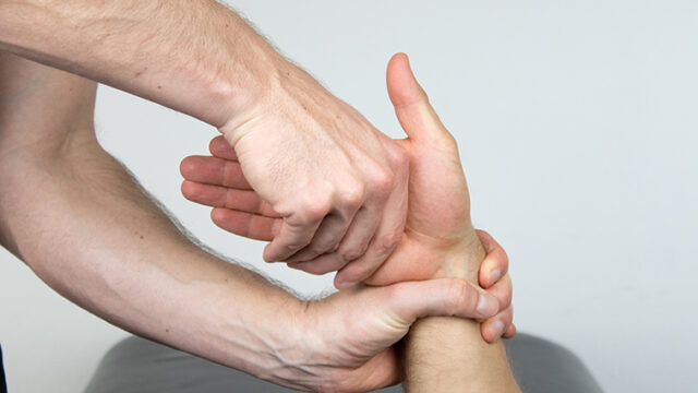
TFCC compression test

TFCC Injury | Diagnosis & Treatment

The triangular fibrocartilage complex (TFCC) sits on the ulnar side of the wrist. A TFCC injury, often called a TFCC tear, is a common source of ulnar-sided wrist pain (Ahn et al., 2006). Many symptomatic TFCC tears can be managed without surgery, especially when the distal radioulnar joint is stable (Choi et al., 2024). Diagnosis can be challenging because clinical tests have limitations, imaging accuracy varies by technique and tear location, and TFCC abnormalities also occur in people without symptoms.

Епидемиология

TFCC injuries are encountered in athletes and people whose activities involve repeated wrist loading or forearm rotation, although reliable incidence estimates are limited. (Liu et al., 2026). Imaging findings are not specific. In 103 asymptomatic volunteers, the TFCC looked abnormal on MRI in 39 wrists, including 23 complete tears, and all three volunteers over 60 years of age had abnormal scans (Iordache et al., 2012).

Mechanism of injury

Both traumatic and degenerative causes occur. They can produce vague ulnar-sided pain, particularly during forearm rotation (Jawed et al., 2020). Early management should consider the injury mechanism, symptom severity, and DRUJ stability. A recent narrative review proposes different early rehabilitation approaches for acute traumatic and repetitive overuse-related presentations. (Liu et al., 2026).

Relevant anatomy

The TFCC stabilises the distal radioulnar joint (DRUJ) and acts as a shock absorber across the ulnocarpal joint. It includes the fibrocartilage disc, the dorsal and palmar radioulnar ligaments, the ulnocarpal ligaments, the meniscal homolog, and the extensor carpi ulnaris tendon subsheath (Jawed et al., 2020).

Pathophysiology and classification

The Palmer classification separates traumatic lesions, classified by location, from degenerative lesions, classified by location and severity (Palmer, 1989). Class 1 covers traumatic tears (1A central, 1B ulnar peripheral, 1C distal, and 1D radial). Class 2 covers degenerative lesions, which relate to ulnar-sided load on the wrist.

Palmer 1B tears vary a lot. A treatment-oriented classification splits them into five classes. Class 1 is an isolated distal TFCC tear without DRUJ instability. Class 2 involves both the distal TFCC and its proximal foveal attachment, whereas Class 3 involves an isolated proximal foveal tear. Both typically produce a positive arthroscopic hook test and are associated with DRUJ instability. Class 4 tears are irreparable. Class 5 tears occur with DRUJ arthritis (Atzei et al., 2017).


Palmer classification of TFCC tears

Клинична презентация и преглед

Injury mechanisms and associated factors
  • Wrist-loading and rotational sports such as racquet sports, golf, gymnastics and combat sports (Liu et al., 2026)
  • Fall on the wrist or other acute trauma
  • Repetitive loading and increasing age (degenerative tears)
  • Positive ulnar variance, which increases ulnocarpal loading and is associated with degenerative TFCC changes.
Клинична картина

Patients report ulnar-sided wrist pain, often worse with forearm rotation, gripping, and weight-bearing on the hand. Clicking, clunking, or a feeling of instability can occur. After trauma, ask about the mechanism, swelling, and any DRUJ symptoms.

Изпит

A systematic, anatomy-based exam of the ulnar wrist is key. Select examination maneuvers according to the suspected structure. These include tests for TFCC-related pain, DRUJ instability, ulnocarpal impaction, and ECU tendon pathology. Provocative tests include the piano key, ulnar impaction, shuck, foveal stress, and extensor carpi ulnaris synergy tests (DaSilva et al., 2017). On our site, see the TFCC Compression Test, Fovea Sign, Ulnar Grinding Test, and DRUJ Test.

Do not rely on a single test. In a retrospective study of 908 patients undergoing arthroscopy for suspected TFCC lesions, the clinical tests evaluated had positive predictive values of 0.53–0.55. The ulnar grinding test had the highest sensitivity but the lowest specificity. The ulnar fovea sign and MRI both had sensitivity of 0.73 to 0.76 and specificity of 0.41 to 0.44 (Schmauss et al., 2016). Combine history, several tests and your clinical reasoning, and always compare with the other wrist.

Diagnosis and imaging

Radiographs help to exclude fractures and look at ulnar variance. A 2012 meta-analysis found that MR arthrography detected full-thickness TFCC tears more accurately than conventional MRI, with pooled sensitivity and specificity of 0.84 and 0.95 compared with 0.75 and 0.81. Data on partial-thickness tears were insufficient (Smith et al., 2012). Because abnormal TFCC findings are common in people without symptoms, interpret imaging together with the history and exam (Iordache et al., 2012). Wrist arthroscopy is increasingly used for diagnosis and treatment of ulnar-sided intra-articular pathology (DaSilva et al., 2017).

Диференциални диагнози

The differential of ulnar-sided wrist pain includes injuries to the ulnar carpal bones, other ligament tears, tendinitis, vascular conditions, osteoarthritis, systemic arthritis and ulnar nerve compression (DaSilva et al., 2017). Our masterclass Ulna-Sided Wrist Pain: A Case Study of a Golfer walks through a clinical reasoning example.

Red flags and referral

Refer early when you see frank DRUJ instability, a suspected fracture (distal radius, ulnar styloid or carpal bones), a locked wrist, or a large traumatic effusion. DRUJ subluxation and full tears of the foveal attachment are associated with poor functional outcome or the need for surgery after nonoperative treatment (Choi et al., 2024). Refer for hand-specialist assessment when pain or functional limitation persists despite an appropriate trial of nonsurgical care, or earlier if DRUJ instability is present.

Лечение

Conservative or surgical?

Asymptomatic injuries do not need treatment. For symptomatic tears with a stable DRUJ, nonsurgical care is usually the first step. Marked instability or associated unstable fractures warrant early specialist assessment. In athletes, all 10 studies of one systematic review used conservative management for 6 weeks to 6 months as first-line treatment, and surgery was advised if symptoms persisted. When surgery was indicated, the review generally recommended arthroscopic debridement for central tears and repair for repairable peripheral tears (Robertson et al., 2019).

A systematic review of eight studies involving 425 patients reported favourable nonsurgical outcomes, particularly in clinically stable injuries. Outcomes appeared similar to those reported in surgical literature, but the limited, heterogeneous evidence does not establish treatment equivalence. For Palmer 1B tears, a short period of above-elbow immobilisation was an effective option. DRUJ subluxation, short-arm immobilisation and full tears of the foveal attachment were risk factors for poor outcome. The authors also say that randomised controlled trials are lacking (Choi et al., 2024).

Surgical results are good but not perfect. In a review of Palmer type 1 injuries, 92% of patients returned to work after debridement but only 44% were pain-free. After repair of 1B lesions, 68.3% returned to work and 41% had persistent pain. These percentages come from different reporting subsets and tear groups, rather than a direct comparison of debridement with repair (McNamara et al., 2020). A systematic review of repair of traumatic 1B tears found improvement in all functional outcomes (for example DASH around 15 to 16 and grip strength 80 to 93% of the other side). Arthroscopic repair had better overall reported outcomes than open repair in this review, but the predominantly low-level evidence does not establish that arthroscopy is superior (Liu et al., 2021).

Initial management

Early management usually involves modification of activities that provoke pain, avoiding sports and gym movements that stress the wrist, and a temporary splint or cast, sometimes with NSAIDs or a corticosteroid injection alongside physical therapy (Jawed et al., 2020). Shock wave therapy and platelet-rich plasma remain investigational for TFCC injuries because condition-specific evidence is limited and does not establish routine benefit (Liu et al., 2026).

Physiotherapy management

Standardised non-surgical rehabilitation pathways remain underdeveloped. One narrative review proposes a phased approach for TFCC injuries without DRUJ instability, with presentation-specific early management, progressive tissue loading and functional reintegration (Liu et al., 2026). The four phases below are a practical adaptation of these principles, not a validated TFCC rehabilitation protocol. Progression should be individualised. Check the stability of the DRUJ first, and follow the surgeon’s protocol after repair.

  • Phase 1, protection and symptom management (first 2 to 6 weeks, depending on the injury pattern, stability and clinical response): relative rest from the provoking activity, a wrist or forearm splint if needed, pain-free range of motion for the fingers, elbow and shoulder, and avoidance of loaded forearm rotation and ulnar deviation under load.
  • Phase 2, range of motion and motor control: restore pain-free wrist flexion, extension and forearm rotation, add isometric and light isotonic work for the wrist flexors and extensors, and train proprioception with closed chain tasks as tolerated.
  • Phase 3, progressive loading: build grip and forearm rotation strength, including the muscles that help to stabilise the DRUJ and ulnar wrist. Increase load gradually and use pain during and after loading to guide progression.
  • Phase 4, return to sport or work: sport-specific or job-specific loading, such as swing, throwing, push-up or weight-bearing tasks. Return when loaded rotation is pain-free and strength is close to the other side. Guide return by symptom response, functional range of motion, strength and tolerance of progressively demanding sport- or work-specific tasks. These are practical considerations rather than validated TFCC return-to-sport criteria.

If there is no meaningful improvement after about 3 to 6 months of well-dosed conservative care, discuss surgical referral. Consider hand-specialist referral when pain or functional limitation persists despite an appropriate nonsurgical trial. Refer earlier for DRUJ instability; the timing should reflect the injury and clinical progress (Robertson et al., 2019).

Return to sport and prognosis

Prognosis is mixed and depends on the tear type and DRUJ stability. Nonsurgical care can produce favourable outcomes in selected patients, but comparative evidence against surgery remains limited (Choi et al., 2024). A substantial proportion of operated patients still have pain (McNamara et al., 2020). Set expectations early and monitor symptoms with a patient-reported outcome measure.

Препратки

Ahn AK, Chang D, Plate AM. Triangular fibrocartilage complex tears: a review. Bull NYU Hosp Jt Dis. 2006.

Liu P, Zhou T. Anatomy, aetiology, clinical presentation, classification, diagnosis, and non-surgical management of triangular fibrocartilage complex tears: a narrative review. Front Sports Act Living. 2026.

Iordache SD, Rowan R, Garvin GJ, et al. Prevalence of triangular fibrocartilage complex abnormalities on MRI scans of asymptomatic wrists. J Hand Surg Am. 2012.

Jawed A, Ansari MT, Gupta V. TFCC injuries: How we treat?. J Clin Orthop Trauma. 2020.

Palmer AK. Triangular fibrocartilage complex lesions: a classification. J Hand Surg Am. 1989.

Atzei A, Luchetti R, Garagnani L. Classification of ulnar triangular fibrocartilage complex tears. A treatment algorithm for Palmer type IB tears. J Hand Surg Eur Vol. 2017.

DaSilva MF, Goodman AD, Gil JA, et al. Evaluation of Ulnar-sided Wrist Pain. J Am Acad Orthop Surg. 2017.

Schmauss D, Pöhlmann S, Lohmeyer JA, et al. Clinical tests and magnetic resonance imaging have limited diagnostic value for triangular fibrocartilaginous complex lesions. Arch Orthop Trauma Surg. 2016.

Smith TO, Drew B, Toms AP, et al. Diagnostic accuracy of magnetic resonance imaging and magnetic resonance arthrography for triangular fibrocartilaginous complex injury: a systematic review and meta-analysis. J Bone Joint Surg Am. 2012.

Choi SI, Malik S, MacLean S. The Natural History of Non-operatively Treated Traumatic Triangular Fibrocartilage Complex Tears: A Systematic Review. J Wrist Surg. 2024.

Robertson G, Ang KK, Maffulli N, et al. Return to sport following surgical management of triangular fibrocartilage tears: a systematic review. Br Med Bull. 2019.

McNamara CT, Colakoglu S, Iorio ML. A Systematic Review and Analysis of Palmer Type I Triangular Fibrocartilage Complex Injuries: Outcomes of Treatment. J Hand Microsurg. 2020.

Liu EH, Suen K, Tham SK, et al. Surgical Repair of Triangular Fibrocartilage Complex Tears: A Systematic Review. J Wrist Surg. 2021.

Искате ли да овладеете горния крайник?

Гледайте безплатен мастърклас на Andrew Cuff за предписване на упражнения при болка в рамото, последван от втори мастърклас на Thomas Mitchell за болка в китката.

  • Това поле е за целите на валидирането и трябва да остане непроменено.
Безплатен мастърклас за горния крайник

REVIEW NOTES (delete this block before publishing)

Open points from the draft for the clinical review. All PubMed references were checked automatically (exist, not retracted, first author and year match); the clinical content still needs checking.

  • Palmer subtypes (1A central, 1B ulnar peripheral, 1C distal, 1D radial; class 2 degenerative) are written from the classic classification. Palmer 1989’s abstract does not list them. Please verify the wording.
  • Risk factors list (positive ulnar variance, fall on the wrist, rotational sports) is partly clinical knowledge; only the athlete and wrist-loading population is cited (Liu 2026). No incidence figure was found.
  • Choi 2024 lists ‘short-arm immobilisation’ as a risk factor for poor outcome and recommends a short period of above-elbow immobilisation for Palmer 1B. This is an unusual recommendation for the clinical reviewer to confirm. The page does not give a duration beyond ‘short period’ and ‘first 2 to 6 weeks’ for the protective phase, which is pragmatic.
  • Rehabilitation phases, time windows and return criteria (e.g. ‘strength close to the other side’) are pragmatic and not from a trial. Liu 2026 is a narrative review and its framework was only read in abstract.
  • Red flag list (locked wrist, large traumatic effusion, suspected fracture) is expert practice and not cited individually.
  • Schmauss 2016 is a retrospective study in patients already scheduled for arthroscopy, with selection bias. Reviewer: consider how strongly to word ‘do not rely on a single test’.
  • Clicking, clunking and instability as symptoms, plus ‘worse with forearm rotation and weight bearing’, are clinical knowledge (Jawed mentions rotation only).
  • Liu 2021 grip strength ’80 to 93%’ is peripheral 80.3 and foveal 92.7%; DASH 15.7 and 15.8. Rounded in text.
  • No NSAID or injection dosing given on purpose. The page mentions injections only as reported in Jawed 2020.
  • No relevant study.physiotutors.com course found, cta_course_url empty.